Review
. 2022 Jul;3(7):793-807.
doi: 10.1038/s43018-022-00411-z. Epub 2022 Jul 26.

Cancer-associated fibroblasts in the single-cell era

Affiliations
Review

Cancer-associated fibroblasts in the single-cell era

Dor Lavie et al. Nat Cancer. 2022 Jul.

Abstract

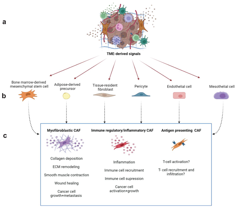
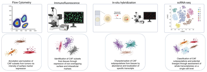
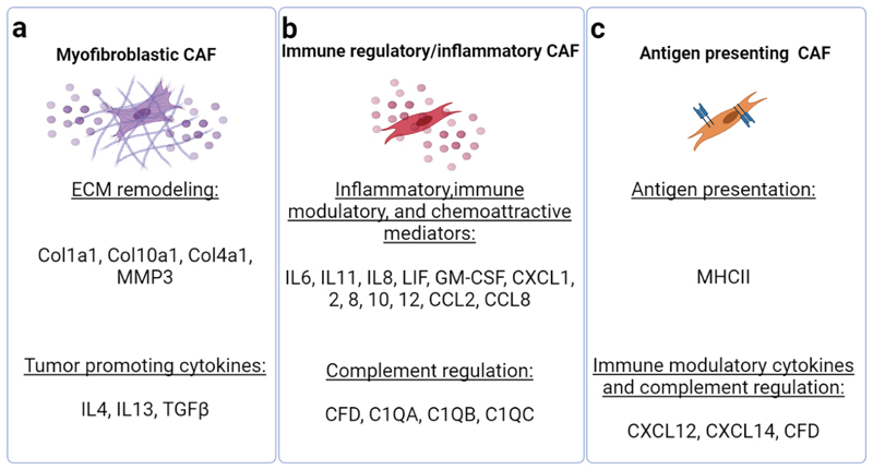
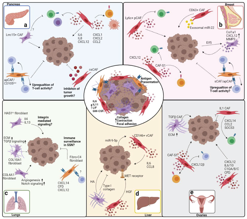
Cancer-associated fibroblasts (CAFs) are central players in the microenvironment of solid tumors, affecting cancer progression and metastasis. CAFs have diverse phenotypes, origins and functions and consist of distinct subpopulations. Recent progress in single-cell RNA-sequencing technologies has enabled detailed characterization of the complexity and heterogeneity of CAF subpopulations in multiple tumor types. In this Review, we discuss the current understanding of CAF subsets and functions as elucidated by single-cell technologies, their functional plasticity, and their emergent shared and organ-specific features that could potentially be harnessed to design better therapeutic strategies for cancer.


Conflict of interest statement

Competing interest statement

The authors declare no competing interests.

Figures

Figure 1
Figure 1. Primary CAF subsets and their potential origins.
TME-derived signals (a) can reprogram a variety of proximal and distal healthy cells, including bone marrow-derived mesenchymal cells, adipocytes, resident fibroblasts, pericytes, endothelial cells, and mesothelial cells (b) into CAFs. The major underlying CAF subgroups can be segregated into myofibroblastic CAFs, immune regulatory/inflammatory CAFs, and antigen presenting CAFs (c) based on the tasks they undertake in the TME, namely reorganization of the ECM, inflammation and modulation of the immune system, and direct effects on cancer cell proliferation and metastatic spread. The figure was created with BioRender.com.
Figure 2
Figure 2. Identification of discrete CAF subsets achieved through different experimental systems.
From left to right: Flow cytometry has been employed to annotate and sort different CAF subgroups from tumors, also enabling CAF subset isolation for further experimentation into CAF phenotypes and tasks. Immunofluorescence and RNA in-situ hybridization experiments revealed CAF subsets from tissue samples, based on discrete expression of surface and intracellular protein markers and transcripts; these methods also reveal important spatial information regarding CAF subtype localization in the TME. Finally, scRNA sequencing has provided a breakthrough in stratification of a multitude of novel CAF subtypes through high resolution characterization of whole transcriptomes on a single cell level, providing information on rare CAF subsets and potential information regarding CAF lineages. The figure was created with BioRender.com.
Figure 3
Figure 3. scRNA-seq reveals multiple tasks undertaken by discrete CAF subtypes.
The major tasks performed by the three central CAF subtypes-ECM reorganization, immune regulation, and antigen presentation, are mediated through expression of various cell surface receptors and secreted factors that influence tumor progression. The secreted factors and receptors listed in the figure generally vary according to disease type and organ, and are designated accordingly in Figure 4. The figure was created with BioRender.com.
Figure 4
Figure 4. scRNA-seq analyses reveal universal, as well as organ-specific CAF subsets.
The main CAF subsets identified in the TME of most organs are inflammatory CAFs, antigen presenting CAFs, and myfibroblastic CAFs (center circle). Immune regulatory (cytokine and chemokine secretion, crosstalk with immune cells) and myofibroblastic CAF activities (ECM modulation, collagen deposition, contraction and adhesion) are prevalent in all organs and across cancer types. Antigen presenting activities of CAFs were predominantly reported in pancreatic and breast cancers. Organ-specific tasks and pathways identified in CAFs are depicted for pancreas (top left), breast (top right), lungs (bottom left), ovaries (bottom center), and liver (bottom right). Arrows indicate promotion of cancer progression and immune cell recruitment/activation. Inhibitory arrows indicate suppression of cancer progression or immune cell activity. Speculative/unverified pathways are labeled with question marks. The figure was created with BioRender.com.
Figure 5
Figure 5. Examples of potential CAF targeting
a targeting LRCC15+ CAFs with antibodies/antibody drug conjugates such as ABBV-085, in conjunction with conventional checkpoint inhibitor therapy (anti-PD-L1) may synergize to abolish their suppressive effect during immunotherapy ,. b Targeting hedgehog signaling pathways via administration of SMO antagonists such as LDE225 can prevent their pro-tumorigenic activities . Since such treatment promotes accumulation of immune suppressive inflammatory CAFs, dual treatment with checkpoint inhibitors may provide important additive effects. c Enhancing the activity of antigen presenting CAFs and their ability to recruit CD4+ T cells could promote anti-tumor immune activity. d Targeting inflammatory CAFs with IL1R antagonists (e.g. anakinra) and anti-TNFα antibodies could inhibit their immune suppressive effects in the TME . The figure was created with BioRender.com.

References

    1. Liu L, et al. Stromal Myofibroblasts Are Associated with Poor Prognosis in Solid Cancers: A Meta-Analysis of Published Studies. Plos One. 2016;11:e0159947. doi: 10.1371/journal.pone.0159947. - DOI - PMC - PubMed
    1. Sahai E, et al. A framework for advancing our understanding of cancer-associated fibroblasts. Nat Rev Cancer. 2020;20:174–186. doi: 10.1038/s41568-019-0238-1. - DOI - PMC - PubMed
    1. Sharon Y, et al. Tumor-derived osteopontin reprograms normal mammary fibroblasts to promote inflammation and tumor growth in breast cancer. Cancer Res. 2015;75:963–973. doi: 10.1158/0008-5472.CAN-14-1990. - DOI - PubMed
    1. Kalluri R. The biology and function of fibroblasts in cancer. Nat Rev Cancer. 2016;16:582–598. doi: 10.1038/nrc.2016.73. - DOI - PubMed
    1. Quante M, et al. Bone marrow-derived myofibroblasts contribute to the mesenchymal stem cell niche and promote tumor growth. Cancer Cell. 2011;19:257–272.:S1535-6108(11)00042-0 [pii] doi: 10.1016/j.ccr.2011.01.020. - DOI - PMC - PubMed

Publication types

MeSH terms