. 2022 Jul 26;19(1):51.
doi: 10.1186/s12989-022-00493-8.

Label-free detection and quantification of ultrafine particulate matter in lung and heart of mouse and evaluation of tissue injury

Affiliations

Label-free detection and quantification of ultrafine particulate matter in lung and heart of mouse and evaluation of tissue injury

Saira Hameed et al. Part Fibre Toxicol. .

Abstract

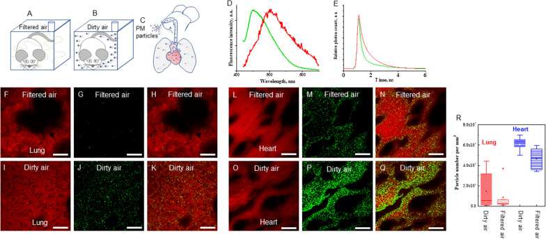
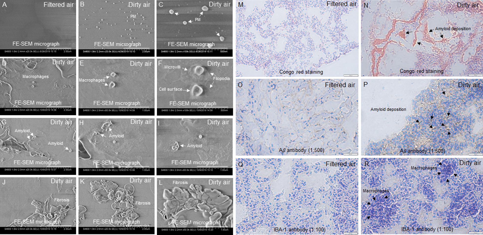
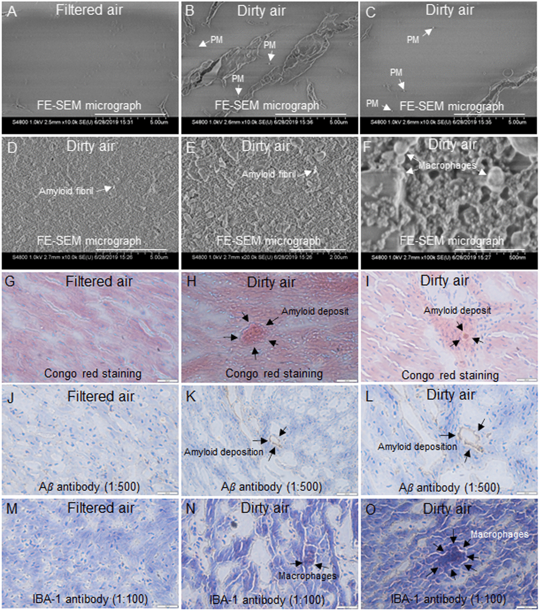
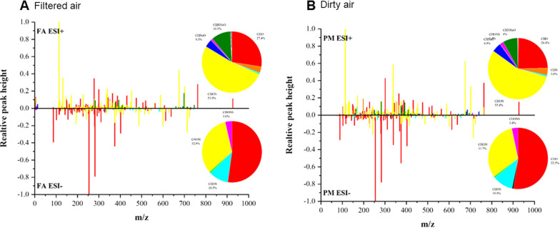
While it is known that air borne ultrafine particulate matter (PM) may pass through the pulmonary circulation of blood at the alveolar level between lung and heart and cross the air-blood barrier, the mechanism and effects are not completely clear. In this study the imaging method fluorescence lifetime imaging microscopy is adopted for visualization with high spatial resolution and quantification of ultrafine PM particles in mouse lung and heart tissues. The results showed that the median numbers of particles in lung of mice exposed to ultrafine particulate matter of diameter less than 2.5 µm was about 2.0 times more than that in the filtered air (FA)-treated mice, and about 1.3 times more in heart of ultrafine PM-treated mice than in FA-treated mice. Interestingly, ultrafine PM particles were more abundant in heart than lung, likely due to how ultrafine PM particles are cleared by phagocytosis and transport via circulation from lungs. Moreover, heart tissues showed inflammation and amyloid deposition. The component analysis of concentrated airborne ultrafine PM particles suggested traffic exhausts and industrial emissions as predominant sources. Our results suggest association of ultrafine PM exposure to chronic lung and heart tissue injuries. The current study supports the contention that industrial air pollution is one of the causative factors for rising levels of chronic pulmonary and cardiac diseases.

Keywords: Fluorescence lifetime imaging microscopy; Heart; Injury; Lung; Scanning electron microscopy; Ultrafine PM.


Conflict of interest statement

The authors declare no competing interests.

Figures

Fig. 1
Fig. 1
Mice were kept in exposure chambers for six months. Graphical illustrations A) Mouse in filtered air chamber. B) Mouse in dirty air chamber. C) Movement of ultrafine PM particles into the respiratory track and accumulation in lungs and heart. D) Normalized fluorescence spectra of ultrafine PM in PBS (green) and auto-fluorescence of tissues (red). E) Typical lifetime decay curves of ultrafine PM in tissues (green) and auto-fluorescence of tissues (red). F) Auto-fluorescent image of lung tissue from filtered air. G) Ultrafine PM particles from filtered air. H) Fluorescent deposition pattern of ultrafine PM particles in lung tissue from filtered air. I) Auto-fluorescent image of lung tissue from dirty air. J) Ultrafine PM particles from dirty air. K) Fluorescent deposition pattern of ultrafine PM particles in lung tissue from dirty air. L) Auto-fluorescent image of heart tissue from filtered air. M) Green dots are the ultrafine PM particles. N) Fluorescent deposition pattern of ultrafine PM particles in heart tissue from filtered air. O) Auto-fluorescent image of heart tissue from dirty air. P) Green dots are the ultrafine PM particles. Q) Fluorescent deposition pattern of ultrafine PM particles in heart tissue from dirty air. Scale bar: 20 µm. The resolution of FLIM images is approximately 250 nm. R) The estimated particle density in lung and heart of mice
Fig. 2
Fig. 2
Field emission scanning electron microscopy (FE-SEM) of lung tissues. A) Lung tissues from filtered air showed no signs of abnormality, whereas B, C lung tissue sections from dirty air exposure group showed ultrafine PM particles, DF macrophages. High magnification FE-SEM image of the macrophages showed cell surface, knob like microvilli, and filopodia that extended outwards from periphery of the cells (F). Lung tissue sections from dirty air exposure group showed amyloid deposits (GI), and fibrosis (JL) Congo red staining, M lung tissues from filtered air (control) showed no signs of abnormality, whereas N lung tissue from the dirty air showed amyloid deposition. Immunohistochemistry with amyloid marker Aβ antibody (1:500), O from lung tissue sections from filtered air (control) group showed no signs of abnormality, whereas P lung tissue from dirty air showed immunoreactive areas with dark brown amyloid deposits. Immunohistochemistry with macrophage marker IBA-1 antibody (1:100), Q lung tissues from filtered air showed no signs of abnormality, whereas R lung tissue from dirty air showed macrophages. Magnification (A, B, D, G, J, and K) 20 k, scale bar: 2 µm. Magnification (E, H, I, and L) 50 k, Scale bar: 1 µm. Magnification (C and F) 100 k, Scale bar: 500 nm. Magnification (M and N) 20X, Scale bar: 500 µm. Magnification (O, P, Q, and R) 40X, Scale bar 500 µm
Fig. 3
Fig. 3
Field emission scanning electron microscopy (FE-SEM) of heart tissues. A Heart tissues from filtered air (control) group showed no signs of abnormality, whereas B, C heart tissue from dirty air exposure group showed ultrafine PM particles, D, E amyloid, and F macrophages. Histopathological evaluation by Congo red staining, G heart tissues from filtered air (control) group showed no signs of abnormality, whereas H, I heart tissue from the dirty air showed amyloid deposition. Immunohistochemistry with amyloid marker Aβ antibody (1:500), J from heart of filtered air (control) group showed no signs of abnormality, whereas) heart tissue from dirty air showed immunoreactive areas with dark brown amyloid deposition (K and L). Immunohistochemistry with macrophage marker IBA-1 antibody (1:100), M heart tissues from filtered air (control) group showed no signs of abnormality, whereas (N and O) heart tissue from dirty air showed macrophages. Magnification (A to D) 10 k, scale bar: 5 μm. Magnification (E) 20 k, scale bar 2 μm. Magnification (F) 100 k, scale bar: 500 nm. Magnification (G, H, I, J, L, M, N, and O) 40X, Scale bar: 500 µm. Magnification (K) 20X, Scale bar: 500 µm
Fig. 4
Fig. 4
Mass spectrometry anslyses of ultrafine PM particles

References

    1. Woodward NC, Crow AL, Zhang Y, Epstein S, Hartiala J, Johnson R, et al. Exposure to nanoscale particulate matter from gestation to adulthood impairs metabolic homeostasis in mice. Sci Rep. 2019;9(1):1816. doi: 10.1038/s41598-018-37704-2. - DOI - PMC - PubMed
    1. Al-Kindi SG, Brook RD, Biswal S, Rajagopalan S. Environmental determinants of cardiovascular disease: lessons learned from air pollution. Nat Rev Cardiol. 2020;17(10):656–672. doi: 10.1038/s41569-020-0371-2. - DOI - PMC - PubMed
    1. Li D, Li Y, Li G, Zhang Y, Li J, Chen H. Fluorescent reconstitution on deposition of PM(25) in lung and extrapulmonary organs. Proc Natl Acad Sci USA. 2019;116(7):2488–2493. doi: 10.1073/pnas.1818134116. - DOI - PMC - PubMed
    1. Hameed S, Zhao J, Zare RN. Ambient PM particles reach mouse brain, generate ultrastructural hallmarks of neuroinflammation, and stimulate amyloid deposition, tangles, and plaque formation. Talanta Open. 2020;2:100013. doi: 10.1016/j.talo.2020.100013. - DOI
    1. Baqir M, Roden AC, Moua T. Amyloid in the lung. Semin Respir Crit Care Med. 2020;41(2):299–310. doi: 10.1055/s-0040-1708059. - DOI - PubMed

Publication types