Comment
. 2022 Jul 26;13(1):43.
doi: 10.1186/s13293-022-00448-w.

Sex differences in the human brain: a roadmap for more careful analysis and interpretation of a biological reality

Affiliations
Comment

Sex differences in the human brain: a roadmap for more careful analysis and interpretation of a biological reality

Alex R DeCasien et al. Biol Sex Differ. .

Abstract

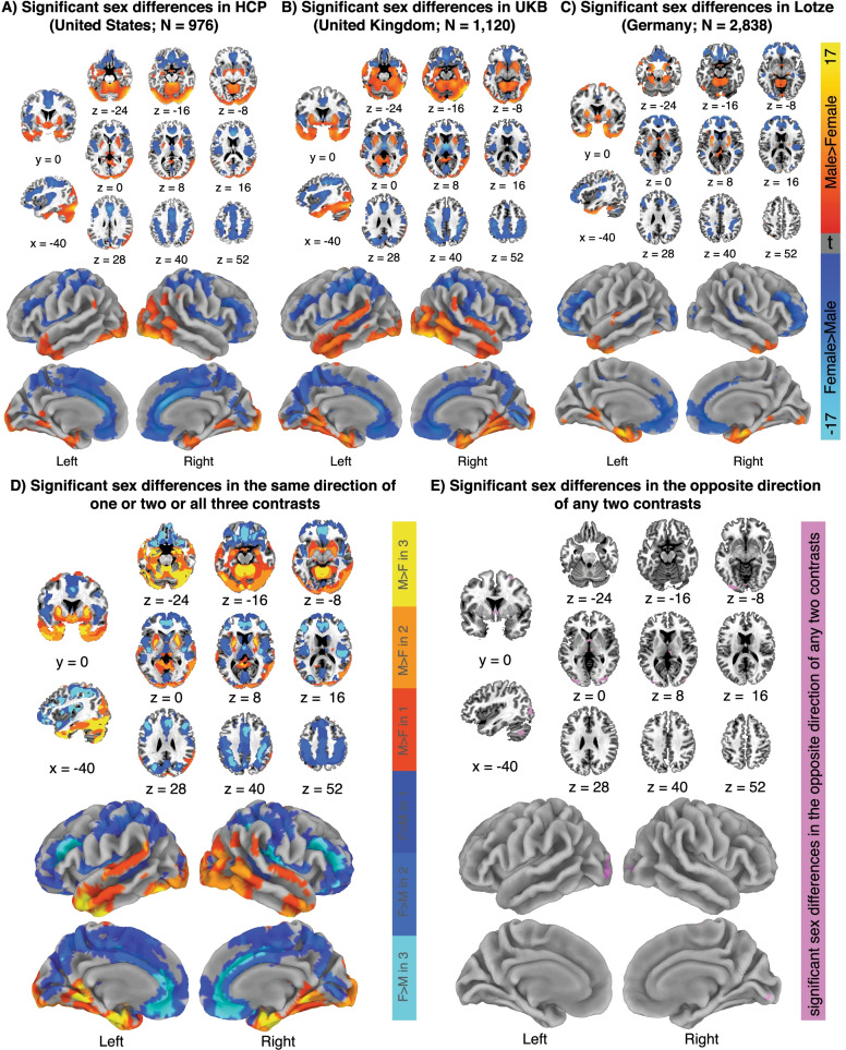
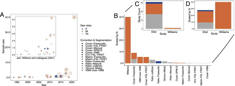
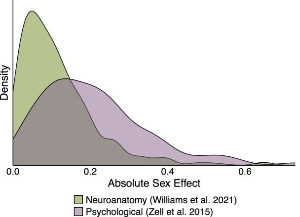
The presence, magnitude, and significance of sex differences in the human brain are hotly debated topics in the scientific community and popular media. This debate is largely fueled by studies containing strong, opposing conclusions: either little to no evidence exists for sex differences in human neuroanatomy, or there are small-to-moderate differences in the size of certain brain regions that are highly reproducible across cohorts (even after controlling for sex differences in average brain size). Our Commentary uses the specific comparison between two recent large-scale studies that adopt these opposing views-namely the review by Eliot and colleagues (2021) and the direct analysis of ~ 40k brains by Williams and colleagues (2021)-in an effort to clarify this controversy and provide a framework for conducting this research. First, we review observations that motivate research on sex differences in human neuroanatomy, including potential causes (evolutionary, genetic, and environmental) and effects (epidemiological and clinical evidence for sex-biased brain disorders). We also summarize methodological and empirical support for using structural MRI to investigate such patterns. Next, we outline how researchers focused on sex differences can better specify their study design (e.g., how sex was defined, if and how brain size was adjusted for) and results (by e.g., distinguishing sexual dimorphisms from sex differences). We then compare the different approaches available for studying sex differences across a large number of individuals: direct analysis, meta-analysis, and review. We stress that reviews do not account for methodological differences across studies, and that this variation explains many of the apparent inconsistencies reported throughout recent reviews (including the work by Eliot and colleagues). For instance, we show that amygdala volume is consistently reported as male-biased in studies with sufficient sample sizes and appropriate methods for brain size correction. In fact, comparing the results from multiple large direct analyses highlights small, highly reproducible sex differences in the volume of many brain regions (controlling for brain size). Finally, we describe best practices for the presentation and interpretation of these findings. Care in interpretation is important for all domains of science, but especially so for research on sex differences in the human brain, given the existence of broad societal gender-biases and a history of biological data being used justify sexist ideas. As such, we urge researchers to discuss their results from simultaneously scientific and anti-sexist viewpoints.

Keywords: Anti-sexism; Direct analysis; Meta-analysis; Neuroanatomy; Neurodevelopment; Review; Sex chromosomes; Sex differences; Sexual selection; sMRI.


Conflict of interest statement

The authors declare that they have no competing interest.

Figures

Fig. 1
Fig. 1
The spatial patterning of neuroanatomical sex differences is largely reproducible across 3 large cohorts. Sex differences in regional gray matter volume (after brain size-correction) across 3 independent cohorts. A Results from the HCP cohort (N = 976) analyzed by Liu and colleagues [8]. B Results from the UKB subsample (N = 1120) analyzed by Liu and colleagues [8]. C Results from the SHIP cohort (N = 2838) analyzed by Lotze and colleagues [9]. D Conjunction map across 3 cohorts. Color encodes whether 1, 2 or 3 of these cohorts show overlapping statistically significant sex differences in regional GMV (cool colors, F > M, warm colors: M > F). Note the spatial nesting of colors (this is consistent with a core pattern of sex biases in regional GMV that is variably recovered by these three studies). E Map of inconsistencies across cohorts. Purple regions are those where any 2 of the 3 cohorts considered showed statistically significant regional GMV sex differences in opposite directions
Fig. 2
Fig. 2
Methodological differences across studies explain apparent inconsistencies in reported sex differences in amygdala volume. Results and design characteristics of studies on sex differences in amygdala volume, including the direct analyses by Williams and colleagues [2] and the studies collated by Eliot and colleagues [1] (N = 31). A Each point represents one study: color = detected sex-bias, size = sample size, shape = brain size correction and segmentation method combination (see legend). Inset depicts the plot excluding Williams and colleagues [2]. B Bar plot depicting the sex-bias (color) per correction and segmentation method (with the Williams et al. study isolated), scaled by the sum of the underlying study sample sizes. C Bar plot depicting the sex-bias (color) per correction and segmentation method, scaled by the underlying study counts. D Bar plot depicting the sex-bias (color) across the studies tallied by Eliot and colleagues and the study by Williams and colleagues, scaled by the sum of the underlying study sample sizes. Loss of information regarding analytical methods and sample sizes accounts for apparently inconsistent findings. Notably, the only studies that detect female-biased amygdala volumes use the proportionalization method for brain size correction (which introduces biases—see text) combined with Freesurfer segmentation. Studies that report non-significant differences tend to have smaller sample sizes (mean N across studies: ns N = 557; female-biased: N = 859; male-biased: N = 4366) or are meta-analyses of studies that used various methods. In fact, all large studies that used the covariate or VBM correction methods (N = 6 studies with sample sizes > 1000) detected male-biased amygdala volumes in their primary analyses
Fig. 3
Fig. 3
Effect size ranges for sex differences in neuroanatomy and psychological measures. Density plot of absolute sex effects (either Cohen’s d or standardized betas) on neuroanatomical and psychological variables. Neuroanatomical sex effects are depicted for 620 neuroanatomical volumes, surface areas, and thicknesses from Williams and colleagues (measured in [2], depicted in [3]). Psychological sex effects were derived from 106 meta-analyses (collated in [137]) across multiple domains, including cognitive (N = 30), personality/social (N = 65), and well-being (N = 11)

Comment on

References

    1. Eliot L, Ahmed A, Khan H, Patel J. Dump the “dimorphism”: comprehensive synthesis of human brain studies reveals few male-female differences beyond size. Neurosci Biobehav Rev. 2021;125:667–697. doi: 10.1016/j.neubiorev.2021.02.026. - DOI - PubMed
    1. Williams CM, Peyre H, Toro R, Ramus F. Neuroanatomical norms in the UK Biobank: the impact of allometric scaling, sex, and age. Hum Brain Mapp. 2021;42:4623–4642. doi: 10.1002/hbm.25572. - DOI - PMC - PubMed
    1. Williams CM, Peyre H, Toro R, Ramus F. Sex differences in the brain are not reduced to differences in body size. Neurosci Biobehav Rev. 2021;130:509–511. doi: 10.1016/j.neubiorev.2021.09.015. - DOI - PubMed
    1. Del Giudice M. Binary thinking about the sex binary: a comment on Joel (2021) Neurosci Biobehav Rev Pergamon. 2021 doi: 10.1016/j.neubiorev.2021.04.020. - DOI - PubMed
    1. Joel D. Beyond the binary: rethinking sex and the brain. Neurosci Biobehav Rev. 2021 doi: 10.1016/j.neubiorev.2020.11.018. - DOI - PubMed