Aquatic Pollution and Risks to Biodiversity: The Example of Cocaine Effects on the Ovaries of Anguilla anguilla
- PMID: 35883315
- PMCID: PMC9312106
- DOI: 10.3390/ani12141766
Aquatic Pollution and Risks to Biodiversity: The Example of Cocaine Effects on the Ovaries of Anguilla anguilla
Abstract
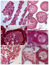
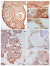
Pollution is one of the main causes of the loss of biodiversity, currently one of the most important environmental problems. Important sources of aquatic pollution are illicit drugs, whose presence in waters is closely related to human consumption; their psychoactive properties and biological activity suggest potential adverse effects on non-target organisms, such as aquatic biota. In this study, we evaluated the effect of an environmentally relevant concentration of cocaine (20 ng L−1), an illicit drug widely found in surface waters, on the ovaries of Anguilla anguilla, a species critically endangered and able to accumulate cocaine in its tissues following chronic exposure. The following parameters were evaluated: (1) the morphology of the ovaries; (2) the presence and distribution of enzymes involved in oogenesis; (3) serum cortisol, FSH, and LH levels. The eels exposed to cocaine showed a smaller follicular area and a higher percentage of connective tissue than controls (p < 0.05), as well as many previtellogenic oocytes compared with controls having numerous fully vitellogenic and early vitellogenic oocytes. In addition, the presence and location of 3β-hydroxysteroid dehydrogenase, 17β-hydroxysteroid dehydrogenase, and P450 aromatase differed in the two groups. Finally, cocaine exposure decreased FSH and LH levels, while it increased cortisol levels. These findings show that even a low environmental concentration of cocaine affects the ovarian morphology and activity of A. anguilla, suggesting a potential impact on reproduction in this species.
Keywords: Anguilla anguilla; cocaine; gonadotropins; oogenesis enzymes; ovary.
Conflict of interest statement
The authors declare that they have no known competing financial interests or personal relationships that could have appeared to influence the work reported in this paper.
Figures


Similar articles
-
Effects of environmental cocaine concentrations on the skeletal muscle of the European eel (Anguilla anguilla).Sci Total Environ. 2018 Nov 1;640-641:862-873. doi: 10.1016/j.scitotenv.2018.05.357. Epub 2018 Jun 5. Sci Total Environ. 2018. PMID: 29879672
-
Changes in the gills of the European eel (Anguilla anguilla) after chronic exposure to environmental cocaine concentration.Ecotoxicol Environ Saf. 2019 Mar;169:112-119. doi: 10.1016/j.ecoenv.2018.11.010. Epub 2018 Nov 13. Ecotoxicol Environ Saf. 2019. PMID: 30445241
-
Effects of environmental cocaine concentrations on COX and caspase-3 activity, GRP-78, ALT, CRP and blood glucose levels in the liver and kidney of the European eel (Anguilla anguilla).Ecotoxicol Environ Saf. 2021 Jan 15;208:111475. doi: 10.1016/j.ecoenv.2020.111475. Epub 2020 Oct 15. Ecotoxicol Environ Saf. 2021. PMID: 33068975
-
Steroidogenic activities of follicle-stimulating hormone in the ovary of Japanese eel, Anguilla japonica.Gen Comp Endocrinol. 2006 Apr;146(2):83-90. doi: 10.1016/j.ygcen.2005.09.019. Epub 2005 Nov 17. Gen Comp Endocrinol. 2006. PMID: 16297918
-
A mechanistic model for studying the initiation of anguillid vitellogenesis by comparing the European eel (Anguilla anguilla) and the shortfinned eel (A. australis).Gen Comp Endocrinol. 2019 Aug 1;279:129-138. doi: 10.1016/j.ygcen.2019.02.018. Epub 2019 Feb 20. Gen Comp Endocrinol. 2019. PMID: 30796898
Cited by
-
Changes of CB1 Receptor Expression in Tissues of Cocaine-Exposed Eels.Animals (Basel). 2025 Jun 12;15(12):1734. doi: 10.3390/ani15121734. Animals (Basel). 2025. PMID: 40564285 Free PMC article.
-
Illicit Drugs in Surface Waters: How to Get Fish off the Addictive Hook.Pharmaceuticals (Basel). 2024 Apr 22;17(4):537. doi: 10.3390/ph17040537. Pharmaceuticals (Basel). 2024. PMID: 38675497 Free PMC article. Review.
-
Cocaine Effects on Reproductive Behavior and Fertility: An Overview.Vet Sci. 2023 Jul 25;10(8):484. doi: 10.3390/vetsci10080484. Vet Sci. 2023. PMID: 37624271 Free PMC article. Review.
References
-
- Murgado-Armenteros E.M., Gutierrez-Salcedo M., Torres-Ruiz F.J. The concern about biodiversity as a criterion for the classification of the sustainable consumer: A cross-cultural approach. Sustainability. 2020;12:3472. doi: 10.3390/su12083472. - DOI
-
- Belpaire C., Goemans G. The European eel Anguilla anguilla, a rapporteur of the chemical status for the water framework directive? Vie Milieu-Life Environ. 2007;57:235–252.
-
- Bettinetti R., Galassi S., Quadroni S., Volta P., Capoccioni F., Ciccotti E., De Leo G. Use of Anguilla anguilla for biomonitoring persistent organic pollutants (POPs) in brackish and riverine waters in central and southern Italy. Water Air Soil Pollut. 2011;217:321–331. doi: 10.1007/s11270-010-0590-y. - DOI
-
- Slingenberg A., Bratt L., van der Windt H., Rademaekers K., Eichler L., Turner K. Study on Understanding the Cause of Biodiversity Loss and the Policy Assessment Framework. European Commission Directorate-General for Environment; Brussels, Belgium: 2009. Contract No.DG.ENV.G.1/FRA/2006/0073 Final Report.
Grants and funding
LinkOut - more resources
Full Text Sources

