. 2022 Jun 30;12(7):922.
doi: 10.3390/biom12070922.

Identification and Characterization of an RRM-Containing, RNA Binding Protein in Acinetobacter baumannii

Affiliations

Identification and Characterization of an RRM-Containing, RNA Binding Protein in Acinetobacter baumannii

Caterina Ciani et al. Biomolecules. .

Abstract

Acinetobacter baumannii is a Gram-negative pathogen, known to acquire resistance to antibiotics used in the clinic. The RNA-binding proteome of this bacterium is poorly characterized, in particular for what concerns the proteins containing RNA Recognition Motif (RRM). Here, we browsed the A. baumannii proteome for homologous proteins to the human HuR(ELAVL1), an RNA binding protein containing three RRMs. We identified a unique locus that we called AB-Elavl, coding for a protein with a single RRM with an average of 34% identity to the first HuR RRM. We also widen the research to the genomes of all the bacteria, finding 227 entries in 12 bacterial phyla. Notably we observed a partial evolutionary divergence between the RNP1 and RNP2 conserved regions present in the prokaryotes in comparison to the metazoan consensus sequence. We checked the expression at the transcript and protein level, cloned the gene and expressed the recombinant protein. The X-ray and NMR structural characterization of the recombinant AB-Elavl revealed that the protein maintained the typical β1α1β2β3α2β4 and three-dimensional organization of eukaryotic RRMs. The biochemical analyses showed that, although the RNP1 and RNP2 show differences, it can bind to AU-rich regions like the human HuR, but with less specificity and lower affinity. Therefore, we identified an RRM-containing RNA-binding protein actually expressed in A. baumannii.

Keywords: Acinetobacter baumannii; ELAVL1; RNA recognition motif.


Conflict of interest statement

The authors declare no conflict of interest.

Figures
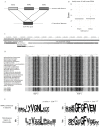
Figure 1
Figure 1
In silico analysis. (A) The tblastn search, using as query the human HuR and restricting the search to A. baumannii genomes, gave 25 hits corresponding to the same orthologous protein which share a high homology with both RRM1 and RRM3. (B) Boxplots showing the percentages of identity with the two RRMs, see Table S1 tblastn for the extended dataset. (C) Genomic context of the bacterial HuR, it is shown that the three genes, namely an ATP-dependent helicase, the AB-Elavl and the ASCH domain containing protein are arranged in proximity. (D) Alignment of the bacterial homologues of human ELAV in selected bacterial species with clinical or environmental relevance spanning seven phyla, along with the HuR RRMs. The background shades denote the level of conservation in that position, darker background mean more conserved residue in that position. (E) Sequence logos for the significantly conserved regions, corresponding to RNP1 and 2 in Prokaryotes (upper row, dataset produced in this study) and Eukaryotes (lower row, dataset from Samson 2008). The seqlogos have been aligned to highlight the presence of conserved residues.
Figure 2
Figure 2
Protein identification and purification. (A) PCR amplification of the transcription of the polycistronic mRNA containing the sequence of interest. The amplicons produced are 390 bp for F1-R2, 340 bp for F2-R1, 764 bp for F3-R3 and 150 bp for F1-R1 (this amplicon was also used as positive control). Neg_ctrl: negative control. (B) Purification of the recombinant protein. FT: flow through, W: wash, EL: elution. (C) Mass spectrometry analysis. The recombinant protein was analyzed at first to confirm the sequence. It was then used as a reference for the analysis of A. baumannii proteome. In red: peptides retrieved with high confidence, underlined: conserved peptides. The predicted molecular weight is 12 KDa for the recombinant protein and 10.8 for the protein from A. baumannii. The predicted isoelectric point is 9.06 for both the proteins. (D) Alignment of AB-Elavl (above) and the RRM3 domain of HuR (below). “|” means that the residues in column are identical.; “:” means that the amino acid in column has been substituted by one with similar characteristics; “.” means that semi-conserved substitutions are observed. (E) Western blot analysis to confirm the presence of the protein of interest in the protein lysate of A. baumannii and in the MCF7 lysate, as well as on the recombinant proteins AB-Elavl and HuR.
Figure 3
Figure 3
Molecular characterization of AB-Elavl. (A) Immunoprecipitation assay on the total protein lysate of A. baumannii. IgG was used as a control. No enrichment of the protein was visible by western blot analysis. (B) Protein ranking based on log2 fold-changes (IP/IgG) for all the proteins identified by MS showing an enrichment of three hypothetical and highly similar RNA-binding proteins (RBPs) in the top ten proteins. (C) Entry code and amino acid sequence of the three hypothetical RBPs based on the IP-MS analysis compared with the rAB-Elavl sequence. Bold retrieved peptides (sequence coverage: 62%, 67% and 91% for D0CAL6, A0A009GG82 and A0A4R5S8D9 proteins, respectively).
Figure 4
Figure 4
AB-Elavl protein structure. (A) Ribbon representation of the three-dimensional structure of the bacterial hypothetical HuR RRM domain. The secondary structural elements and loops have been annotated: helices (α1–α2), strands (β1–β5), loops (L1–L7). (B) Superposition of the crystal structure of the bacterial hypothetical HuR RRM domain (rAB-Elavl, red) and 1FLX (green).
Figure 5
Figure 5
2D 1H-15N HSQC spectrum of AB-Elavl. The spectrum was recorded with a spectrometer operating at 900 MHz and 298 K. Assignment is reported on the signals.
Figure 6
Figure 6
Analysis of the protein binding abilities. (A) REMSA assay on the total protein lysate of A. baumannii and a probe mimicking the AU rich sequence of TNFα (ARE pos) with an infrared tag. (B) REMSA assay on the recombinant protein incubated with different probes with an infrared tag: ARE pos and ARE neg. ARE pos is bound with a high affinity, while ARE neg shows a lower affinity. (C) REMSA assay for detection of the super-shift in presence of the antibody against AB-Elavl. The super-shift is not detectable.
Figure 7
Figure 7
NMR analysis of the protein binding abilities. (A). Plots of decreases in signal intensity of rAB-Elavl RRM domain in the presence of 140 µM ARE pos (top), or 140 µM ARE neg (bottom) with respect to the free protein (70 µM). The residues experiencing the largest decreases have been highlighted in blue. (B). Chemical shift perturbation (CSP) of rAB-Elavl RRM domain (70 µM) with respect to the protein in the presence of 140 µM positive RNA (top), and 140 µM negative RNA (bottom). The CSP was evaluated with the formula: δ=12δH2+δN/52. The residues experiencing the largest CSP have been highlighted in red. (C) Highlighted in blue are the residues experiencing the largest decreases in signal intensity, in red the residues experiencing the largest CSP, and in violet the residues experiencing the largest decreases in signal intensity and CSP, in the presence of 140 µM positive RNA (top), and 140 µM negative RNA (bottom).
Figure 8
Figure 8
Biochemical characterization of the protein binding ability. (A) Sequences of the probes used in the different assays. (B) AlphaScreen saturation experiment between the recombinant protein and AREpos and AREneg. The EC50 was determined from non-linear regression fits of the data according to the dose–response model in GraphPad Prism®, version 6.1. (C) AlphaScreen saturation assay for detection of the minimal probe length for binding of the protein. The probe are AREpos with 3′ deletions: ARE pos: ARE sequence full length, ARE pos 19: ARE sequence with 19 nucleotides, ARE pos 11: ARE sequence with 11 nucleotides. The minimal number of nucleotides in order to obtain the binding is 19, but longer sequences have a higher affinity. The EC50 was determined from nonlinear regression fits of the data according to the dose–response model in GraphPad Prism®, version 6.1. (D) EC50 evaluation through saturation experiment by HTRF-FRET and AREpos and AREneg. AREpos is confirmed to have high affinity while AREneg is not well bound. The EC50 was determined from nonlinear regression fits of the data according to the dose–response model in GraphPad Prism®, version 6.1. (E,F) Kinetic experiment with rAB-Elavl and AREpos (E) is dose dependent, while for AREneg (F) the binding resulted ambiguous. Association (Kon) and dissociation (Koff) rate constants were determined from nonlinear regression fits of the data according to association kinetic model of multiple ligand concentration in GraphPad Prism®, version 6.1.

References

    1. Maris C., Dominguez C., Allain F.H.T. The RNA recognition motif, a plastic RNA-binding platform to regulate post-transcriptional gene expression. FEBS J. 2005;272:2118–2131. doi: 10.1111/j.1742-4658.2005.04653.x. - DOI - PubMed
    1. Samson M.L. Rapid functional diversification in the structurally conserved ELAV family of neuronal RNA binding proteins. BMC Genom. 2008;9:392. doi: 10.1186/1471-2164-9-392. - DOI - PMC - PubMed
    1. Corley M., Burns M.C., Yeo G.W. How RNA-Binding Proteins Interact with RNA: Molecules and Mechanisms. Mol. Cell. 2020;78:9–29. doi: 10.1016/j.molcel.2020.03.011. - DOI - PMC - PubMed
    1. Holmqvist E., Vogel J. RNA-binding proteins in bacteria. Nat. Rev. Microbiol. 2018;16:601–615. doi: 10.1038/s41579-018-0049-5. - DOI - PubMed
    1. Colombrita C., Silani V., Ratti A. ELAV proteins along evolution: Back to the nucleus? Mol. Cell Neurosci. 2013;56:447–455. doi: 10.1016/j.mcn.2013.02.003. - DOI - PubMed

Publication types

LinkOut - more resources