. 2022 Jul 16;12(7):991.
doi: 10.3390/biom12070991.

Cloning and Characterization of Drosophila melanogaster Juvenile Hormone Epoxide Hydrolases (JHEH) and Their Promoters

Affiliations

Cloning and Characterization of Drosophila melanogaster Juvenile Hormone Epoxide Hydrolases (JHEH) and Their Promoters

Dov Borovsky et al. Biomolecules. .

Abstract

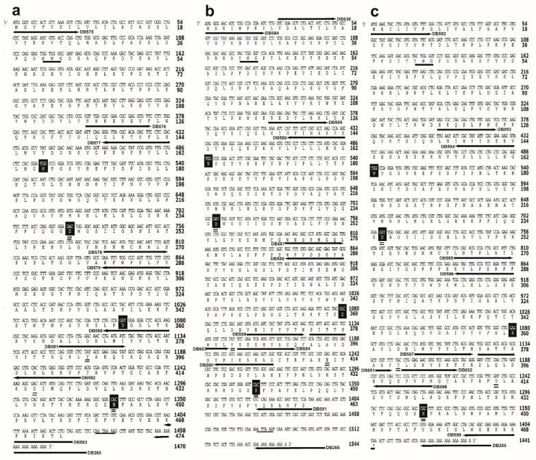
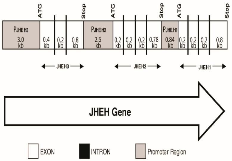
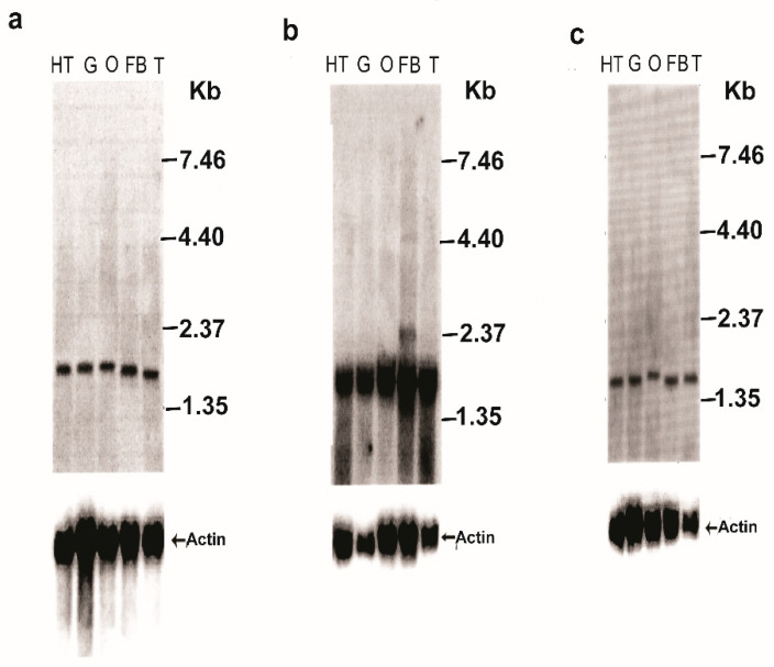
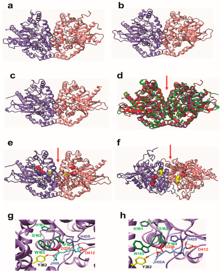
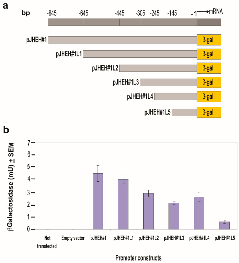
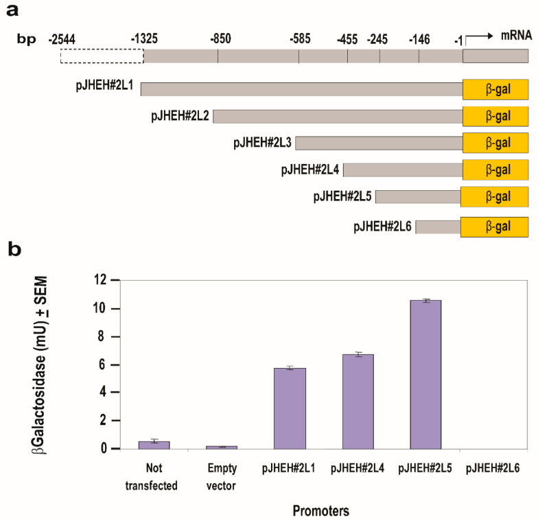
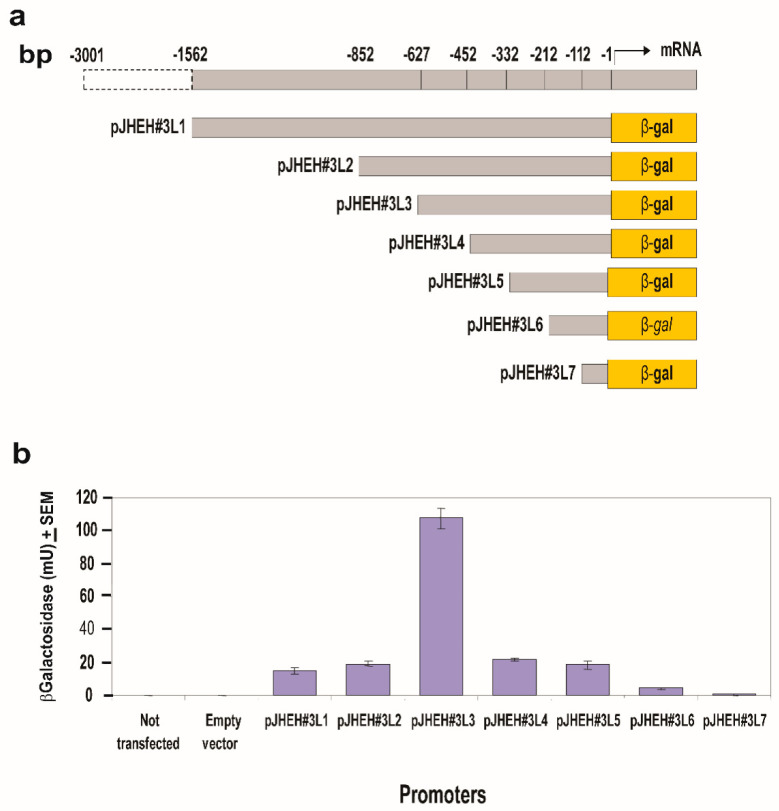
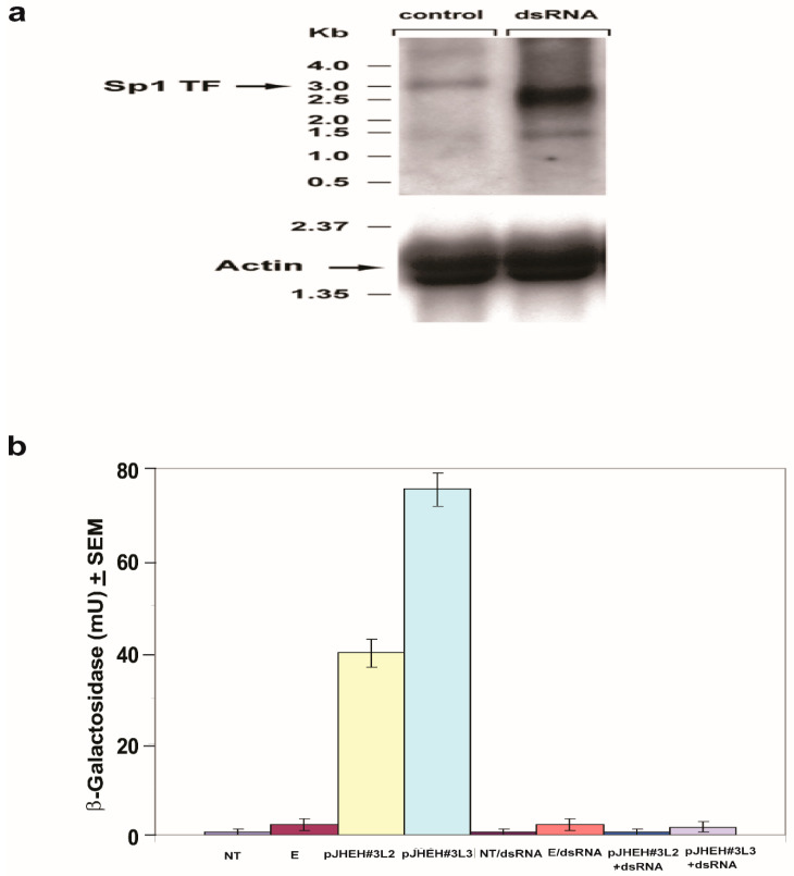
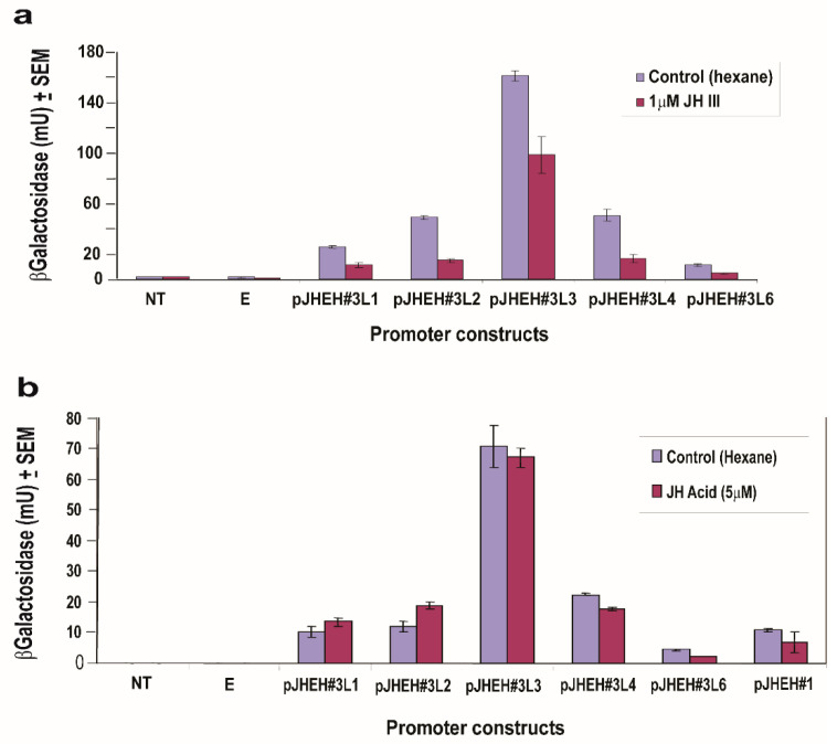
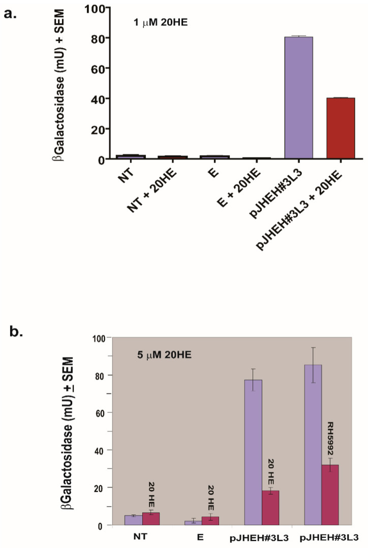
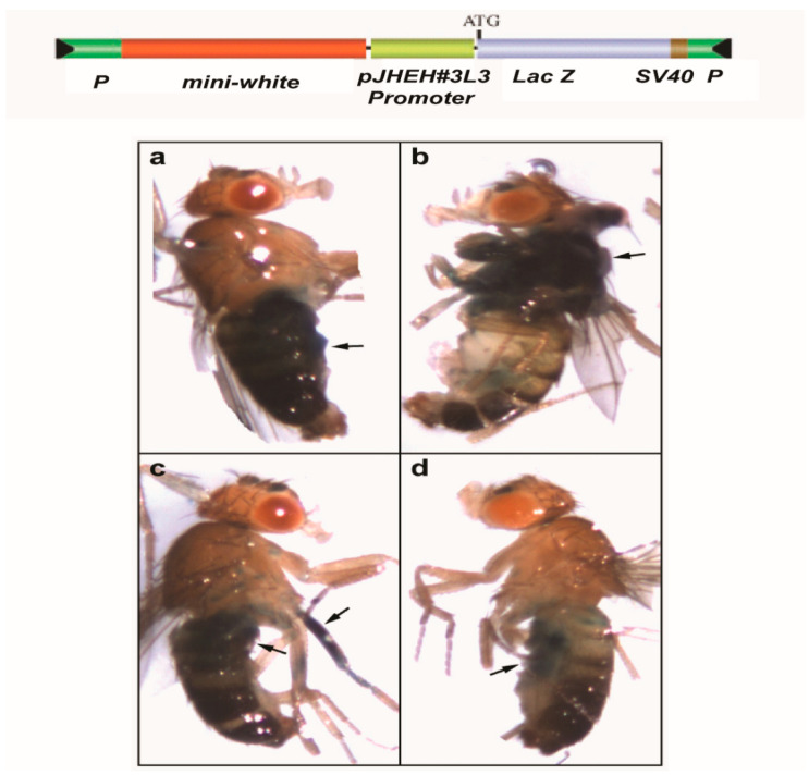
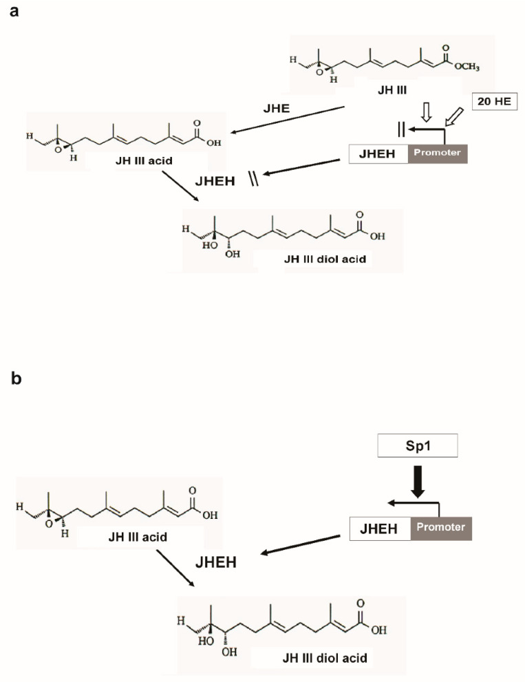
Juvenile hormone epoxide hydrolase (JHEH) plays an important role in the metabolism of JH III in insects. To study the control of JHEH in female Drosophila melanogaster, JHEH 1, 2 and 3 cDNAs were cloned and sequenced. Northern blot analyses showed that the three transcripts are expressed in the head thorax, the gut, the ovaries and the fat body of females. Molecular modeling shows that the enzyme is a homodimer that binds juvenile hormone III acid (JH IIIA) at the catalytic groove better than JH III. Analyses of the three JHEH promoters and expressing short promoter sequences behind a reporter gene (lacZ) in D. melanogaster cell culture identified a JHEH 3 promoter sequence (626 bp) that is 10- and 25-fold more active than the most active promoter sequences of JHEH 2 and JHEH 1, respectively. A transcription factor (TF) Sp1 that is involved in the activation of JHEH 3 promoter sequence was identified. Knocking down Sp1 using dsRNA inhibited the transcriptional activity of this promoter in transfected D. melanogaster cells and JH III and 20HE downregulated the JHEH 3 promoter. On the other hand, JH IIIA and farnesoic acid did not affect the promoter, indicating that JH IIIA is JHEH's preferred substrate. A transgenic D. melanogaster expressing a highly activated JHEH 3 promoter behind a lacZ reporter gene showed promoter transcriptional activity in many D. melanogaster tissues.

Keywords: 3D modeling; RNAi; drosophila; embryo transformation; juvenile hormone epoxide hydrolases promoters; sequencing; tissue culture; transcription factors.


Conflict of interest statement

The authors declare no conflict of interest.

Figures

Figure 8
Figure 8
Testing promoter sequence (225 bp) between pJHEH#3L2 and pJHEH#3L3 (a) for TFs DNA-binding sequences and effects on jheh 3 promoter transcriptional activities, as shown in Figure 7, by testing different promoter lengths and mutating or deleting TF TCF-1 DNA-binding site using primers DB877 and DB876, respectively (b,c).
Figure 1
Figure 1
jheh 1, 2 and 3 cDNA cloning strategies and primers (arrows) that were used in the cloning and the sequencing. (a). jheh 1 cDNA amino acids and nucleotides sequence, the membrane anchor sequence WWG is single underlined, the catalytic amino acids triad DYD is double underlined, the H that participates in the catalytic activity is double underlined and the polyadenylation signal sequence AATAAA is single underlined. (b). jheh 2 cDNA amino acids and nucleotides sequence, the membrane anchor sequence YWG is single underlined, the catalytic amino acids triad DYE is double underlined, the H that participates in the catalytic activity is double underlined and the poly adenylation signal ATTAAA is single underlined. (c). jheh 3 cDNA amino acids and nucleotides sequence, the membrane anchor sequence YWG is single underlined, the catalytic amino acids triad DYD is double underlined, the H that participates in the catalytic activity is double underlined and the poly adenylation signal is not shown.
Figure 2
Figure 2
Genomic DNA sequence of jheh including intron (black lines) exons (white colored squares) and promoters (gray colored squares) of jheh 1, jheh 2 and jheh 3. Arrow follows the 5′ to 3′direction of D. melanogaster genome.
Figure 3
Figure 3
Northern blot analyses of D. melanogaster jheh 1 t (a), jheh 2 (b) and jheh 3 (c) transcripts in the head gut (HT), gut (G), ovary (O), fat body (FB) and total insect extract (T) of female D. melanogaster. Blots were hybridized with specific probes for each transcript (Tables S1–S3). Actin probe was used to show even transfer to the blot. The Northern blot analyses were repeated twice, showing similar results.
Figure 4
Figure 4
3D models showing non-covalently associated homodimeric monomers of (a) JHEH 1 (b) JHEH 2 and (c) JHEH 3. The two monomers are colored violet and pink, respectively. (d) Superpositioning of the 3 models built for JHEH 1 (red), JHEH 2 (blue) and JHEH 3 (green) showing good superposition of the α-helices and β-sheets forming the backbones of the homodimers. The red arrow indicates the depression separating both monomers, associated with the catalytic grooves of both monomers. (e) Lateral view and (f) upper view of JHEH 3 homodimer showing the localization of residues D236 (red), Y382 (yellow), D412 (red) and H439 (blue) forming the catalytic grooves located on either side of the central depression (red arrow). (g). Docking of juvenile hormone III (JH III) (colored cyan) to the catalytic groove of JHEH 3. Amino acid residues D236, D412 and H439 forming the catalytic triad of JHEH 3 are colored red and blue, respectively. Tyrosine residue Y382, forming a H-bond with the epoxide group of JH III, is colored yellow. Amino acid residues from the HGWP motif are colored dark green. Hydrogen bonds are indicated by black dashed lines. (h). Docking of juvenile hormone III acid (JH IIIA) (colored blue), to the catalytic groove of JHEH 3. Amino acid residues D236, D412 and H439 forming the catalytic triad of JHEH 3 are colored red and blue, respectively. Tyrosine residue Y382, forming a H-bond with the epoxide group of JH IIIA, is colored yellow. Amino acid residues from the HGWP motif are colored dark green. Hydrogen bonds are indicated by black dashed lines. Note that only 2 H-bonds out of total 4 H-bonds of D412 with JH IIIA can be seen at this viewing angle.
Figure 5
Figure 5
Testing jheh 1 different promoter sequence lengths (a) for transcriptional activity (b) by cloning them into plasmid pCaSpeR-AUG-βgal, transfecting D.Mel2 cells with the recombinant plasmid and assaying 2 × 10⁵ cells for β-galactosidase activity expressed in milliunits (mU). Controls were D.Mel2 cells that were not transfected or transfected with empty plasmid.
Figure 6
Figure 6
Testing jheh 2 different promoter sequence lengths (a) for transcriptional activity (b) by cloning them into plasmid pCaSpeR-AUG-βgal, transfecting D.Mel2 cells with the recombinant plasmid and assaying 2 × 10⁵ cells for β-galactosidase activity expressed in milliunits (mU). Controls were D.Mel2 cells that were not transfected or transfected with empty plasmid.
Figure 7
Figure 7
Testing jheh 3 different promoter sequence lengths (a) for transcriptional activity (b) by cloning them into plasmid pCaSpeR-AUG-βgal, transfecting D.Mel2 cells with the recombinant plasmid and assaying 2 × 10⁵ cells for β-galactosidase activity expressed in milliunits (mU). Controls were D.Mel2 cells that were not transfected or transfected with empty plasmid.
Figure 9
Figure 9
Northern blot analysis of TF Sp1 in D.Mel2 cells that were transfected with promoter pJHEH#3L3 and treated with dsRNA against Sp1. (a) In cells that were treated with dsRNA, the Sp1 transcript is degraded (right lane) compared with untreated cells (left lane). (b) Transcriptional activity of pJHEH#3L2 and pJHEH#3L3, in transfected D.Mel2 cells is not affected, whereas the activity in cells that were treated with dsRNA is similar to cells that were not transfected (NT) or cells that were transfected with an empty plasmid ©.
Figure 10
Figure 10
Transcriptional activities of different jheh 3 promoter sequences in transfected D.Mel2 cells in the presence of JH III (1 μM) (a) and in the presence of JH IIIA (5 μM) (b). The different promoter sequence lengths are found Figure 8 and Figure 6. Non-transfected cells (NT) and cells transfected with empty plasm © (E).
Figure 11
Figure 11
Effects of 20HE and its mimic (RH5992), 5 μM each, on the transcriptional activity of D.Mel2 transfected with jheh 3 promoter pJHEH#3L3. (a) Red colored bars show incubations with 20HE (1 μM), magenta-colored bars show incubations without 20HE. (b). Red-colored bars show incubations with 20HE or RH5992 (5 μM), magenta-colored bars show incubations without 20HE. NT—Non-transfected cells, E—cells transfected with an empty plasmid. See Figure 7 for promoter pJHEH#3L3 sequence length.
Figure 12
Figure 12
D. melanogaster flies that were transformed with jheh 3 promoter pJHEH#3L3 (colored green) in pCaSpeR-AUG-βgal behind lacZ (colored magenta) upper bar. Arrows point to β-galactosidase activity in the abdomen (a), in the thorax (b), in the leg muscle and upper abdomen next to the thorax (c) and the ventral part of the abdomen (d).
Figure 13
Figure 13
Degradative steps in JH III pathway. (a) JHEH promoter is inhibited by high titers of JH III and 20HE. During this time, JHEH is not synthesized. (b) After JH III is converted into JH IIIA (JH III acid) by JHE, the JHEH promoter is upregulated by TF Sp1, allowing the synthesis of JHEH, which converts JH IIIA into JH IIIAD (JH III acid diol).

References

    1. Riddiford L.M. Cellular and molecular actions of juvenile hormone general considerations and premetamorphic actions. Adv. Insect Physiol. 1994;24:213–273.
    1. Wyatt G.R., Davey K.G. Cellular and molecular actions of juvenile hormone. II. Roles of juvenile hormone in adult insects. Adv. Insect Physiol. 1996;26:1–156.
    1. Stay B., Tobe S.S., Bendena W.G. Allatostatins: Identification, primary structure, function and distribution. Adv. Insect Physiol. 1994;125:267–337.
    1. Hammock B.D., Sparks T.C. A rapid assay for insect juvenile hormone esterase activity. Anal. Biochem. 1977;82:573–579. doi: 10.1016/0003-2697(77)90196-8. - DOI - PubMed
    1. Share M.R., Roe R.M. A partition assay for the simultaneous determination of insect juvenile hormone esterase and epoxide hydrolase activity. Anal. Biochem. 1988;169:81–88. doi: 10.1016/0003-2697(88)90257-6. - DOI - PubMed

Substances

LinkOut - more resources