. 2022 Jul 6;11(14):2129.
doi: 10.3390/cells11142129.

Vascular Remodeling Is a Crucial Event in the Early Phase of Hepatocarcinogenesis in Rodent Models for Liver Tumorigenesis

Affiliations

Vascular Remodeling Is a Crucial Event in the Early Phase of Hepatocarcinogenesis in Rodent Models for Liver Tumorigenesis

Margaret Tulessin et al. Cells. .

Abstract

The investigation of hepatocarcinogenesis is a major field of interest in oncology research and rodent models are commonly used to unravel the pathophysiology of onset and progression of hepatocellular carcinoma. HCC is a highly vascularized tumor and vascular remodeling is one of the hallmarks of tumor progression. To date, only a few detailed data exist about the vasculature and vascular remodeling in rodent models used for hepatocarcinogenesis. In this study, the vasculature of HCC and the preneoplastic foci of alteration (FCA) of different mouse models with varying genetic backgrounds were comprehensively characterized by using immunohistochemistry (CD31, Collagen IV, αSMA, Desmin and LYVE1) and RNA in situ hybridization (VEGF-A). Computational image analysis was performed to evaluate selected parameters including microvessel density, pericyte coverage, vessel size, intratumoral vessel distribution and architecture using the Aperio ImageScope and Definiens software programs. HCC presented with a significantly lower number of vessels, but larger vessel size and increased coverage, leading to a higher degree of maturation, whereas FCA lesions presented with a higher microvessel density and a higher amount of smaller but more immature vessels. Our results clearly demonstrate that vascular remodeling is present and crucial in early stages of experimental hepatocarcinogenesis. In addition, our detailed characterization provides a strong basis for further angiogenesis studies in these experimental models.

Keywords: animal model; foci of cellular alteration; hepatocellular carcinoma; image analysis; vascular remodeling; vessel analysis.


Conflict of interest statement

The authors declare no conflict of interest.

Figures

Figure 1
Figure 1
Annotation and immunostaining of foci of cellular alteration (FCA) and hepatocellular carcinoma (HCC). FCA annotated with green color in H&E (A); HCC annotated with yellow color in H&E (B); representative image of FCA (C) (H&E) and HCC (D) (H&E); CD31 immunostaining in FCA I and HCC (E,F). Collagen IV immunostaining in FCA (G) and HCC (H). Scale bar (A,B): 2 mm (magnification 1.5×). Scale bar (CF): 50 µm (magnification 20×).
Figure 2
Figure 2
Detailed vessel analysis by CD31 and Collagen IV in FCA and HCC. Total number of vessels in FCA versus HCC by CD31 (A) and Collagen IV (B); average staining intensity of vessels by CD31 (C) and Collagen IV (D). No differences were observed in total vessel area: CD31 (E) and Collagen IV (F). Error bars show the mean and standard deviation for each lesion. p-values: Not statistically significant (ns) p value > 0.05; for statistical significance, accepted *** = p value ≤ 0.001, and **** = p value ≤ 0.0001.
Figure 3
Figure 3
Computer-assisted subgrouping of vessels. CD31-based vessel subgrouping (AD) for FCA (A) and CD31 (B) subgroups and HCC (C) and CD31 (D) subgroups. Collagen IV-based vessel subgrouping (EH) for FI (E) and Collagen IV (F) subgroups and HCC (G) and Collagen IV (H) subgroups. Arrows mark the lesion in (AF). Magnification 5×. Color coding of subgroups: (A,D) black-colored areas highlight small-sized vessels (<150 µm2), yellow-colored areas highlight medium-sized vessels (150–500 µm2), and grey-colored areas highlight large-sized vessels (>500 µm²). (F,H) Grey-colored areas highlight small-sized vessels (<150 µm2), yellow-colored areas highlight medium-sized vessels (150–500 µm2), and black-colored areas highlight large-sized vessels (>500 µm2).
Figure 4
Figure 4
Analysis of vessel area and vessel number per lesion. Evaluation of subgrouped vessels per total area in CD31 (AC) and Collagen IV staining (D,E,F). Subgrouped vessels per area in CD31 (GI) and Collagen IV staining (J,K,L). Error bars show the mean and standard deviation for each lesion. p-values: not statistically significant (ns) p value > 0.05; for statistical significance, accepted p value ≤ 0.05., ** p value ≤ 0.01, *** p value ≤ 0.001, and **** p value ≤ 0.0001.
Figure 5
Figure 5
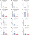
Analysis of staining intensity per lesion of CD31- and Collagen IV-stained vessels (A–F). Error bars indicate the mean and standard deviation for each lesion. p-values: Not statistically significant (ns) p value > 0.05; for statistical significance, accepted p value ≤ 0.05. ** p value ≤ 0.01, *** p value ≤ 0.001, and **** p value ≤ 0.0001.
Figure 6
Figure 6
Heatmap of vessel distribution according to size. Annotated FCA (encircled green) and HCC (encircled yellow) in CD31 staining. (A) Distribution of the vessel according to their size in small-sized vessels (B,C), medium-sized vessels (D,E) and large-sized vessels (F,G) show a predominant location of small- and medium-sized vessels in the center (intralesional) of FCA, whereas the small- and medium-sized vessels in HCC mostly located at the periphery of HCC (arrowheads). In Collagen IV (H) small vessels located in the center (intralesional) of FCA but at the periphery of HCC, with shift towards the periphery in medium- and large-sized vessels (IN). Color coding of heatmap: Green color indicates lowest density, yellow color indicates medium density and red color indicates highest density. Scale bar (A,B): 2 mm (magnification 1×). p-values: Not statistically significant (ns) p value > 0.05; for statistical significance, accepted p value ≤ 0.05. ** p value ≤ 0.01, N = number of hotspots identified in each slide and lesion for further in-depth analysis.
Figure 7
Figure 7
Analysis of vessels per lesion for expression of smooth muscle actin (α-SMA). (A) Immunostaining of α-SMA in FCA and HCC, (B) number of α-SMA positive pixel/total area and (C) intensity of α-SMA/positive pixel. for statistical significance, accepted p value: * p value ≤ 0.05. Scale bar: 50 µm (magnification 20×).

References

    1. Ferlay J., Colombet M., Soerjomataram I., Parkin D.M., Pineros M., Znaor A., Bray F. Cancer statistics for the year 2020: An overview. Int. J. Cancer. 2021;149:778–789. doi: 10.1002/ijc.33588. - DOI - PubMed
    1. Ferlay J., Colombet M., Soerjomataram I., Mathers C., Parkin D.M., Pineros M., Znaor A., Bray F. Estimating the global cancer incidence and mortality in 2018: GLOBOCAN sources and methods. Int. J. Cancer. 2019;144:1941–1953. doi: 10.1002/ijc.31937. - DOI - PubMed
    1. Siegel R.L., Miller K.D., Jemal A. Cancer statistics, 2020. CA Cancer J. Clin. 2020;70:7–30. doi: 10.3322/caac.21590. - DOI - PubMed
    1. Sung H., Ferlay J., Siegel R.L., Laversanne M., Soerjomataram I., Jemal A., Bray F. Global Cancer Statistics 2020: GLOBOCAN Estimates of Incidence and Mortality Worldwide for 36 Cancers in 185 Countries. CA Cancer J. Clin. 2021;71:209–249. doi: 10.3322/caac.21660. - DOI - PubMed
    1. Dhamija E., Paul S.B., Kedia S. Non-alcoholic fatty liver disease associated with hepatocellular carcinoma: An increasing concern. Indian J. Med. Res. 2019;149:9–17. doi: 10.4103/ijmr.IJMR_1456_17. - DOI - PMC - PubMed

Publication types