. 2022 Jul 8;11(14):2153.
doi: 10.3390/cells11142153.

Preclinical Development of a Therapy for Chronic Traumatic Spinal Cord Injury in Rats Using Human Wharton's Jelly Mesenchymal Stromal Cells: Proof of Concept and Regulatory Compliance

Affiliations

Preclinical Development of a Therapy for Chronic Traumatic Spinal Cord Injury in Rats Using Human Wharton's Jelly Mesenchymal Stromal Cells: Proof of Concept and Regulatory Compliance

Joaquim Vives et al. Cells. .

Abstract

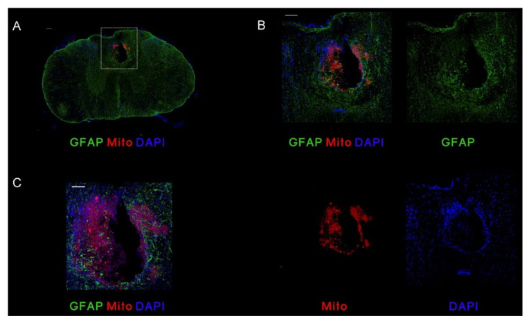
(1) Background: the use of Mesenchymal Stromal Cells (MSC) in emerging therapies for spinal cord injury (SCI) hold the potential to improve functional recovery. However, the development of cell-based medicines is challenging and preclinical studies addressing quality, safety and efficacy must be conducted prior to clinical testing; (2) Methods: herein we present (i) the characterization of the quality attributes of MSC from the Wharton's jelly (WJ) of the umbilical cord, (ii) safety of intrathecal infusion in a 3-month subchronic toxicity assessment study, and (iii) efficacy in a rat SCI model by controlled impaction (100 kdynes) after single (day 7 post-injury) and repeated dose of 1 × 10⁶ MSC,WJ (days 7 and 14 post-injury) with 70-day monitoring by electrophysiological testing, motor function assessment and histology evaluation; (3) Results: no toxicity associated to MSC,WJ infusion was observed. Regarding efficacy, recovery of locomotion was promoted at early time points. Persistence of MSC,WJ was detected early after administration (day 2 post-injection) but not at days 14 and 63 post-injection. (4) Conclusions: the safety profile and signs of efficacy substantiate the suitability of the presented data for inclusion in the Investigational Medicinal Product Dossier for further consideration by the competent Regulatory Authority to proceed with clinical trials.

Keywords: advanced therapy; animal model; cell therapy; cell-based therapy; good laboratory practice; mesenchymal stromal cells; preclinical research; spinal cord injury; stem cells; translational medicine.


Conflict of interest statement

The authors declare no conflict of interest.

Figures

Figure 1
Figure 1
Growth profiles of MSC cultures in presence or absence of FK506. The growth of MSC,WJ in the two experimental settings is presented as the increase of ATP content, measured by luminescence.
Figure 2
Figure 2
Immunopotency assay for MSC in presence or absence of FK506. Both conditions tested resulted in pronounced inhibition of the proliferation of stimulated NC in culture. Values of inhibition of proliferation in co-cultures of MSC,WJ:NC were normalized to proliferating stimulated NC in absence of MSC,WJ.
Figure 3
Figure 3
Effects of MSC,WJ administration on functional recovery. Open-field locomotion was evaluated weekly after spinal cord injury (SCI) using the score (A) and subscore (B) of the Basso, Beattie, Bresnahan (BBB) scale in the three studied groups (each group n = 5): vehicle or VHC (group of animals with SCI and vehicle administered intrathecally at 7 days post-lesion, or dpl), MSC,WJ 7 dpl (group of animals with SCI and 1 × 10⁶ MSC,WJ cells intrathecally at 7 dpl, arrows) and MSC,WJ 7 + 14 dpl (group of animals with SCI and 1 × 10⁶ MSC,WJ cells intrathecally at 7 and 14 dpl, arrows). All animals showed a temporal paralysis (BBB score of 0) just after the injury with partial recovery until day 14 and a plateau phase until the end of follow-up. * p < 0.05 group MSC,WJ 7 + 14 vs. VHC.
Figure 4
Figure 4
Motor nerve conduction studies at 70 dpi. (A) Amplitude of M and H waves recorded in the plantar muscles. The H wave was significantly reduced in MSC,WJ 7 + 14 days post-lesion (dpl) vs. vehicle (VHC). (B) Spinal reflex H/M ratio of plantar muscle showed a significant decrease in MSC,WJ 7 + 14 dpl group vs. VHC. (* p < 0.05). n = 5 per group.
Figure 5
Figure 5
MSC,WJ localization after intrathecal administration at 9 days post-lesion (dpl) and 2 days post-injection (dpi). Representative images of rats with transplanted MSC,WJ. Cells were injected intrathecally (L3–L4). After 2 days, staining for Mito was observed inside the lesion site. (A) Representative microphotography of transversal section showing injured spinal cord at 9 dpl after 2 dpi intrathecal injection of MSC,WJ. (B) Magnification of injured spinal cord (from white box on A) showing GFAP staining in green, Mito staining in red and 4′,6-diamidino-2-phenylindole (DAPI) staining in blue. MSC,WJ can be identified into the lesion site. (C) Confocal microphotography shows localization of engrafted cells at lesion site. Bar scale: 100 µm. n = 3.
Figure 6
Figure 6
MSC,WJ localization after intrathecal administration at 21 or 70 days post-lesion (dpl), 14 days post-injection (dpi) or 63/56 + 63 dpi). Representative images of each group with transplanted MSC,WJ. No staining for SC101 was observed around or inside the lesion. GFAP staining in red, SC101 in green and DAPI in blue. Bar scale: 100 µm. n = 3.
Figure 7
Figure 7
Histological studies. (A) Representative longitudinal images from each experimental condition. From left to right: Glial fibrillary acidic protein (GFAP) staining in red (marked astrocytic reactivity around the lesion), Iba1 staining in red (immune cell recruited around the injury), and RT-97 staining in green (axonal pathways within the injured spinal cord). Bar scale: 100 µm. (B) Scheme of measured areas in B. Quantification of estimated % volume of spared tissue. Significant increase of spared tissue was observed in MSC,WJ 7 + 14 dpl group vs. vehicle (VHC); * p < 0.05. (C) Scheme of measured areas in C. Measurements of immunolabeled density of microglia (Iba1), astroglial reactivity (GFAP) and axonal reactivity rostral, caudal and lesion site (epicenter) at 70 days post-lesion (dpl) did not reveal changes induced by MSC,WJ administration cells. Scale bar: 100 μm. n = 5 in each condition.

References

    1. Simpson L.A., Eng J.J., Hsieh J.T., Wolfe D.L., Team SCIRESR The health and life priorities of individuals with spinal cord injury: A systematic review. J. Neurotrauma. 2012;29:1548–1555. doi: 10.1089/neu.2011.2226. - DOI - PMC - PubMed
    1. Kim Y.-H., Ha K.-Y., Kim S.-I. Spinal Cord Injury and Related Clinical Trials. Clin. Orthop. Surg. 2017;9:1–9. doi: 10.4055/cios.2017.9.1.1. - DOI - PMC - PubMed
    1. Cofano F., Boido M., Monticelli M., Zenga F., Ducati A., Vercelli A., Garbossa D. Mesenchymal Stem Cells for Spinal Cord Injury: Current Options Limitations, and Future of Cell Therapy. Int. J. Mol. Sci. 2019;20:2698. doi: 10.3390/ijms20112698. - DOI - PMC - PubMed
    1. Assinck P., Duncan G.J., Hilton B.J., Plemel J.R., Tetzlaff W. Cell transplantation therapy for spinal cord injury. Nat. Neurosci. 2017;20:637–647. doi: 10.1038/nn.4541. - DOI - PubMed
    1. Naji A., Eitoku M., Favier B., Deschaseaux F., Rouas-Freiss N., Suganuma N. Biological functions of mesenchymal stem cells and clinical implications. Cell. Mol. Life Sci. 2019;76:3323–3348. doi: 10.1007/s00018-019-03125-1. - DOI - PMC - PubMed

Publication types