Review
. 2022 Jul 16;11(14):2216.
doi: 10.3390/cells11142216.

BMP Signaling Pathway in Dentin Development and Diseases

Affiliations
Review

BMP Signaling Pathway in Dentin Development and Diseases

Mengmeng Liu et al. Cells. .

Abstract

BMP signaling plays an important role in dentin development. BMPs and antagonists regulate odontoblast differentiation and downstream gene expression via canonical Smad and non-canonical Smad signaling pathways. The interaction of BMPs with their receptors leads to the formation of complexes and the transduction of signals to the canonical Smad signaling pathway (for example, BMP ligands, receptors, and Smads) and the non-canonical Smad signaling pathway (for example, MAPKs, p38, Erk, JNK, and PI3K/Akt) to regulate dental mesenchymal stem cell/progenitor proliferation and differentiation during dentin development and homeostasis. Both the canonical Smad and non-canonical Smad signaling pathways converge at transcription factors, such as Dlx3, Osx, Runx2, and others, to promote the differentiation of dental pulp mesenchymal cells into odontoblasts and downregulated gene expressions, such as those of DSPP and DMP1. Dysregulated BMP signaling causes a number of tooth disorders in humans. Mutation or knockout of BMP signaling-associated genes in mice results in dentin defects which enable a better understanding of the BMP signaling networks underlying odontoblast differentiation and dentin formation. This review summarizes the recent advances in our understanding of BMP signaling in odontoblast differentiation and dentin formation. It includes discussion of the expression of BMPs, their receptors, and the implicated downstream genes during dentinogenesis. In addition, the structures of BMPs, BMP receptors, antagonists, and dysregulation of BMP signaling pathways associated with dentin defects are described.

Keywords: BMP receptors; Smads; bone morphogenetic proteins (BMPs); canonical Smad signaling; dentin; dentin defects; downstream genes; non-canonical Smad signaling; odontoblasts.


Conflict of interest statement

The authors declare no conflict of interest.

Figures
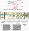
Figure 1
Figure 1
Tooth development. (a) The crown of the tooth is covered with enamel, while the root is covered with cementum and periodontal ligament. The cementoenamel junction is located between the enamel and root. The root is surrounded by the alveolar bone through periodontal ligaments. The dentin surrounds the dental pulp. Nerves, lymph, and blood vessels enter the dental pulp from the apical foramen of the tooth and provide nutrition and innervation to odontoblasts and dental pulp. (b) Teeth form from the surface epithelium and underlying mesenchyme, and their development is regulated by interactions between the dental epithelium and mesenchyme. The early stages are morphologically similar as the ectodermal placodes develop, form buds, and induce the formation of the dental papilla. In the tooth germ, epithelial folding is regulated by the enamel knot signaling center and it determines the shape of the tooth. During tooth development, it goes through various stages of tooth initiation: bud, cap, bell, and eruption. (c) Dentin consists of inter-tubular dentin and dentinal tubules. The figure was partially adapted with permission from Thesleff, 2003, ref. [4].
Figure 2
Figure 2
Phylogenetic analysis of the BMP family. Relationship between BMP/GDF ligands, type I receptors, type II receptors, Smad proteins, and RGM in signal transduction. ActRIIA/B, activin type II receptor A or B; ALK, activin receptor-like kinase; BMP, bone morphogenic protein; BMPRI, BMP type receptor I; BMPRII, BMP type receptor II; GDF, growth differentiation factor; MSTN, myostatin; OP, osteogenic protein; RGM, repulsive guidance molecules; Vgr, Vg-related protein. The figure was partially adapted with permission from Katagiri and Watabe, 2016, ref. [22].
Figure 3
Figure 3
Bmp-2 expression during mouse tooth development. At the cap stage (E15) of mouse tooth development, Bmp-2 mRNA was highly expressed in the dental epithelium and moderately detectable in dental mesenchymal cells, osteogenetic mesenchyme, and Meckel’s cartilage. At the early bell stage (E16), the Bmp-2 transcript was present in ameloblasts, odontoblasts, dental pulp cells, dental follicles, and mesenchyme in the alveolar bone. At postnatal days ranging from 1 to 14, an in situ hybridization signal was apparent in odontoblasts, dental pulp cells, and osteoblasts in the alveolar bone and was weakly detected in ameloblasts. The figure is adapted with permission from Chen et al., 2008, ref. [5]. ab, alveolar bone; am, ameloblasts; mc, Meckel’s cartilage; D, dentin; de, dental epithelium; df, dental follicle; dm, dental mesenchyme; dp, dental papilla; E, enamel; E, embryonic day; od, odontoblasts.
Figure 4
Figure 4
Diagram representing the structure of dimeric BMPs. (a) The BMP-2 monomer fold represents the α-helices and β-sheets shown by spiral and arrows, respectively. The BMP-2 cystine knot constitutes the core of the monomer and consists of three disulfide bonds (“S”); two, Cys-325-Cys-393 and Cys-329-Cys-325, form a ring through which the third, Cys-296-Cys-361, passes. The strands of antiparallel β-sheets of β1 to β9, which emit from the knot, form two fingerlike projections. An α-helix, located on the opposite end of the knot, lies perpendicular to the axis of the two fingers, thus forming the heel of the hand. The N terminus corresponds to the thumb of the hand (adapted from Carreira et al., 2014, ref. [21]). (b) The dimeric structure of BMP-2. Each monomer is indicated by one color (green and orange), with the disulfide bridges being denoted by projections of the cysteine (yellow) side chains (adapted with permission from Scheufler et al., 1999., ref. [125]). The color figure is available online at https://www.rcsb.org/structure/3BMP, accessed on 15 December 2021. (c,d) Crystal structure of BMP-3 and BMP-6 in pink and green (adapted with permission from Allendorph et al., 2007, ref. [128]; online at https://www.rcsb.org/structure, accessed on 17 June 2022). 2QCQ and 2QCW. (e) Crystal structure of BMP-7 (adapted with permission from Griffith et al., 1996, ref. [124]; online at https://www.rcsb.org/structure/1BMP, accessed on 17 June 2022).
Figure 5
Figure 5
Crystal structure of BMPs and receptors. (a) Architecture of the interaction of BMP-2 and ALK-3. Stereoview of the structure of the BMP-2 (green and magenta) and ALK-3 (blue and pink) complex. BCSB.org; PDB ID code 1 REW (adapted with permission from Keller et al., 2004; ref. [132]). (b) The ternary complex of BMP-2–ALK-3 ECD–ActRII-ECD displays a butterfly-like conformation. BMP-2 dimer subunits are shown in red and orange; the two ALK-3-ECDs are indicated in light blue and blue, and the ActR-II-ECDs are shown in light green and green. BCSB.org; PDB ID code 2GOO (adapted with permission from Allendorph et al., 2006, ref. [133]). (c) The crystal structure of the BMP-2 and RGMb complex. Cartoon representation of the BMP-2 and RGMbND complex. BMP2 is shown in blue and magenta; RGMCND is shown in blue. RCSB.org; PDB ID code 4UHY (adapted with permission from Healey et al., 2015; ref. [134]).
Figure 6
Figure 6
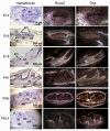
Runx2 and Osx expression patterns in developing teeth. Runx2 mRNA was clearly detected in dental and osteogenic mesenchyme from E14 to E16. Its expression was intense in osteogenic mesenchymal cells, whereas expression was downregulated in odontoblasts, ameloblasts, and dental pulp cells at E18 of the mandibular incisor and first molar. At this stage, Osx expression mostly overlapped with Runx2 expression from E14–E16. At E18, its expression was prominent in ameloblasts, odontoblasts, and dental pulp cells. From PN 1 to PN 14, Runx2 mRNA was detected in osteogenic mesenchyme and remarkedly downregulated in ameloblasts, odontoblasts, and dental pulp cells. In contrast, Osx expression continued to be present in osteogenic mesenchyme, odontoblasts, dental pulp cells, and periodontal ligament cells. ab, alveolar bone; am, ameloblasts; D, dentin; de, dental epithelium; df, dental follicle; dp, dental papilla; E, enamel; E, embryonic day; mc, mesenchymal condensates; od, odontoblasts; pdl, periodontal ligament cells. PN, postnatal day. Photos are partially adapted with permission from Chen et al., 2009, ref. [143].
Figure 7
Figure 7
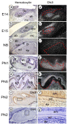
Expression of Dlx3 during mouse tooth development. (ae). Bright images. (a’e’) In situ hybridization for Dlx3 expression during mouse tooth formation. At the cap stage (E14–E15), the anti-Dlx3 probe is strongly present in the dental mesenchyme and osteogenetic mesenchyme developing alveolar bone and is moderately visible in the dental epithelium and Meckel’s cartilage. At new birth (NB) and PNs 1 and 5, expression of Dlx3 can be detected in ameloblasts, odontoblasts, dental papilla, and dental follicles, as well as in the mesenchyme in the alveolar bone. At PN 1, Dlx3 expression is more intense in ameloblasts, odontoblasts, and dental papilla than in developing alveolar bone mesenchymal cells. (f,f’). Tooth tissues were immunostained with anti-Dlx3 antibody, showing that the Dlx3 expression is apparent in ameloblasts, dental pulp cells, and odontoblasts at PN 2 of mouse tooth development. (f’) shows higher a magnification of the box in (f). The tissue sections were stained with anti-IgG serum as the negative control. (g’) shows a higher magnification of the box in (g). ab, alveolar bone; am, ameloblasts; con, control; D, dentin; de, dental epithelium; dm, dental mesenchyme; df, dental follicle; dp, dental papilla; E, enamel; E, embryonic day; mc, Meckel’s cartilage; od, odontoblasts; PN, postnatal day. The data are unpublished.
Figure 8
Figure 8
Dmp1 and Dspp expression patterns in developing teeth. By in situ hybridization assay, Dmp1 mRNA transcription was clearly detected in odontoblasts near the dental crown and root of the mouse tooth and osteoblasts in alveolar bones by PN 1, while by PN 5, Dmp1 expression was downregulated in odontoblasts and only appeared in odontoblasts located at the dental root but was highly intense in the alveolar bone. By PN 10–14, Dmp1 transcription was weakly visible in odontoblasts, but still highly expressed in osteoblasts in alveolar bones. By PN 1, Dspp transcripts were evident in odontoblasts, dental papilla, and pre-ameloblasts in the mouse mandibular molars. By PN 5, Dspp mRNA was strongly expressed in odontoblasts and weakly seen in ameloblasts and dental pulp cells. From PN 10 to 14, Dspp mRNA was mainly expressed in odontoblasts and weakly in dental pulp cells. Dspp expression was barely detected in osteoblasts in alveolar bone from PN 1 to PN 14 during mouse tooth development. ab, alveolar bone; am, ameloblasts; D, dentin; df, dental follicle; dp, dental papilla; E, enamel; od, odontoblasts; mc, Meckel’s cartilage; pAm, pre-ameloblasts; pd, dental pulp; PN, postnatal day. The photos are partially adapted with permission from Chen et al., 2009, ref. [143].
Figure 9
Figure 9
BMP ligands, receptors, and interacting receptors. BMP signal transduction involves a number of ligands, type I and type II serine/threonine kinase receptors, and coreceptors, which regulate the activation of intracellular mediators in interactions with extracellular stimuli. BMPs are classified into several subfamilies according to sequence homology. The BMP signaling pathways. Canonical Smad signaling executes its function through Smad proteins. BMPs interact with the type I and type II receptors to form a heterotetrameric complex. Complex formation and ligand binding can be potentiated by a coreceptor, that is, endoglin, betaglycan, or repulsive guidance molecule (RGM). Upon phosphorylation by the type II receptor, the type I receptor recruits and phosphorylates pathway-specific R Smads (Smad-1, Smad-5 and Smad-8, and Smad-2 and Smad-3), which can form trimers with Smad-4 and translocate to the nucleus. Smads have intrinsic DNA-binding activity and can regulate gene expression by recruitment of chromatin-remodeling machinery and integration with tissue-specific transcription factors. Non-canonical Smad signaling, through phosphorylated TAK (pTAK). Ligand binding induces the formation of the receptor complex. BmpR-I is linked to the TAK1/TAB1 complex through the X-linked inhibitor of apoptosis (XIAP) to phosphorylate and activate the downstream mitogen-activated protein kinases (MAPKs), including p38, ERK1/2, and JNK. Consequently, activation of these kinases leads to their translocation into the nucleus where they phosphorylate and activate and regulate the transcription of downstream target genes. Canonical Smad signaling is intracellularly inhibited by inhibitory Smads, that is, Smad-6 and/or Smad-7, and E3 ubiquitin ligases, such as Smurf1 or Smurf2, whose expression provides the cell with a negative feedback mechanism. The pathway can be antagonized by many mechanisms, including neutralization of ligands by secreted traps, such as noggin, follistatin, gremlin, chordin, chordin-like; secretion of latent ligands bound to their propeptides; or via titration of receptors by nonsignaling ligands, such as BMP-3. ACVR, activin receptor; ACVR2, activin type-2 receptor; ALK, activin receptor-like kinase; AMH, anti-Müllerian hormone; AMHR2, AMH receptor; BMP, bone morphogenetic protein; BmpR, bone morphogenetic protein receptor; ERK, mitogen-activated protein kinase; GDF, growth/differentiation factor; JNK, c-Jun N-terminal kinase; MAPK, mitogen-activated protein kinase; p38, p38 mitogen-activated protein kinases; RGM, repulsive guidance molecule; SBE, Smad binding element; TAK, TGF-activated kinase; TF, transcriptional factor; TGF, transforming growth factor; TGFBR, TGF-β receptor; XIAP, E3 ubiquitin-protein ligase XIAP. The figure is partially adapted with permission from Miyazone et al., 2005, ref. [279].
Figure 10
Figure 10
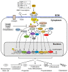
Smad-dependent signaling in dentin formation. BMPs bind to receptor type II (R-II) and receptor type I (R-I) before the signaling transduces to their Smad1/5/8. Activated Smad1/5/8 interacts with Smad4, forming a complex which translocates into the nucleus where they interact with other transcription factors to trigger target gene expression. Consequently, Smad-dependent signaling enhances dental mesenchymal cell differentiation and expression of DSPP and DMP1 genes via the action of Smads, Dlx3, Dlx5, Klf4, Msx1, Msx2, Osx, Pax9, and Runx2, and other compounds during dentinogenesis. Smad6 binds the type I BMP receptor and prevents Smad1/5/8 from being activated. Additionally, Smurf1 interacts with the Runx2 protein and induces Runx2 protein degradation. The figure is partially adapted with permission from Wu et al., 2016, ref. [291].
Figure 11
Figure 11
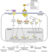
Smad-independent signaling pathways. BMP signal transduction involves a number of ligands, type I and type II serine/threonine kinase receptors and coreceptors, which regulate the activation of intracellular mediators in interactions with extracellular stimuli. Smad-independent signaling through phosphorylated TAK (pTAK), protein kinases A and C (PKA/C), and PI3K/Akt. Ligand binding induces the formation of the receptor complex. BMP receptors linked to the TAK1/TAB1 phosphorylate and activate the downstream MAPKs, including p38, ERK1/2, and JNK, respectively. Consequently, activation of these kinases leads to the phosphorylation of several transcriptional factors, activating the transcription of downstream target genes. In addition, PI3K/Akt and PKA/C are phosphorylated by BMP receptors. Then activated PI3K/Akt and PKA/C phosphorylate their downstream transcription factors. These activated transcription factors bind to their binding responsible elements in their target gene regulatory regions and upregulate the expression of DSPP and DMP1 genes and induce dental mesenchymal cell differentiation and dentin formation. Akt, (v-Akt murine thymoma viral oncogene)/PKB (protein kinase B); ERKs, extracellular signal-regulated kinases; JNKs, c-Jun NH2-terminal kinases; PI3K, phosphoinositol 3-Kinase; PKA, protein kinase A; PKC, protein kinase C. The figure is partially adapted with permission from Wu et al., 2016, ref. [291].
Figure 12
Figure 12
BMP signaling is inhibited by BMP antagonists during tooth development. The antagonists include noggin, gremlin, chordin, chordin-like 1, chordin-like 2, and others. Noggin interacts with BMPs or Wnt ligands and prevents the binding of BMPs to their receptors and Wnt-Frizzled receptors, consequently blocking their signaling and impairing dentin development and formation. Gremlin and chordin, chordin-like 1, and chordin-like 2 also bind to BMPs and interfere with dentinogenesis via canonical Smad and non-Smad signaling pathways. →, activation; ┤, inhibition. Wnt, wingless type MMTV integration site family; SBE, Smad binding element; BMP, bone morphogenetic protein; MAPK, mitogen-activated protein kinase; p38, p38 mitogen-activated protein kinases; ERK, mitogen-activated protein kinase; TF, transcriptional factor; Akt, v-Akt murine thymoma viral oncogene/PKB (protein kinase B). Figure is partially adapted with permission from Katagiri et al., 2016, ref. [22].

References

    1. Mitsiadis T.A., Orsini G., Jimenez-Rojo L. Stem cell-based approaches in dentistry. Eur. Cell Mater. 2015;30:248–257. doi: 10.22203/eCM.v030a17. - DOI - PubMed
    1. Nanci A. Ten Cate’s Oral Histology: Development, Structure, and Function. 8th ed. Mosby; St. Louis, MI, USA: 2012. pp. 1–13.
    1. Zhang Y.D., Chen Z., Song Y.Q., Liu C., Chen Y.P. Making a tooth: Growth factors, transcription factors, and stem cells. Cell Res. 2005;15:301–316. doi: 10.1038/sj.cr.7290299. - DOI - PubMed
    1. Thesleff I. Epithelial-mesenchymal signaling regulating tooth morphogenesis. J. Cell Sci. 2003;116:1647–1648. doi: 10.1242/jcs.00410. - DOI - PubMed
    1. Chen S., Gluhak-Heinrich J., Martinez M., Li T., Wu Y., Chuang H.H., Chen L., Dong J., Gay I., MacDougall M. Bone morphogenetic protein 2 mediates dentin sialophosphoprotein expression and odontoblast differentiation via NF-Y signaling. J. Biol. Chem. 2008;283:19359–19370. doi: 10.1074/jbc.M709492200. - DOI - PMC - PubMed

Publication types

Substances