. 2022 Jun 29;11(7):1294.
doi: 10.3390/antiox11071294.

Next-Generation Sequencing Advances the Genetic Diagnosis of Cerebral Cavernous Malformation (CCM)

Affiliations

Next-Generation Sequencing Advances the Genetic Diagnosis of Cerebral Cavernous Malformation (CCM)

Valerio Benedetti et al. Antioxidants (Basel). .

Abstract

Cerebral Cavernous Malformation (CCM) is a cerebrovascular disease of genetic origin that predisposes to seizures, focal neurological deficits and fatal intracerebral hemorrhage. It may occur sporadically or in familial forms, segregating as an autosomal dominant condition with incomplete penetrance and highly variable expressivity. Its pathogenesis has been associated with loss-of-function mutations in three genes, namely KRIT1 (CCM1), CCM2 and PDCD10 (CCM3), which are implicated in defense mechanisms against oxidative stress and inflammation. Herein, we screened 21 Italian CCM cases using clinical exome sequencing and found six cases (~29%) with pathogenic variants in CCM genes, including a large 145−256 kb genomic deletion spanning the KRIT1 gene and flanking regions, and the KRIT1 c.1664C>T variant, which we demonstrated to activate a donor splice site in exon 16. The segregation of this cryptic splicing mutation was studied in a large Italian family (five affected and seven unaffected cases), and showed a largely heterogeneous clinical presentation, suggesting the implication of genetic modifiers. Moreover, by analyzing ad hoc gene panels, including a virtual panel of 23 cerebrovascular disease-related genes (Cerebro panel), we found two variants in NOTCH3 and PTEN genes, which could contribute to the abnormal oxidative stress and inflammatory responses to date implicated in CCM disease pathogenesis.

Keywords: KRIT1/CCM1; aberrant splicing; cerebral cavernous malformation (CCM); cerebrovascular disease; clinical exome sequencing (CES); genetic modifiers; next-generation sequencing (NGS).


Conflict of interest statement

The authors declare no conflict of interest.

Figures
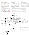
Figure 1
Figure 1
Sanger validation and pedigrees. (A) Electropherograms of the identified CCM gene variants reported in Table 2. (B) Four-generation pedigree of family 12. Filled symbols indicate affected subjects; a question mark indicates the subjects carrying the pathogenic variant but without signs of disease. The genotype is indicated below each tested subject (wt/wt, homozygous wild type; wt/+, heterozygote for the KRIT1 c.1664C>T variant). (C) Pedigree of family 1 (left) and electropherogram of the identified NOTCH3 c.2960C>G variant (right).
Figure 2
Figure 2
Analysis of the KRIT1 genomic deletion identified by NGS in case #7. (A) Screenshot from UCSC genome browser centered on KRIT1 (hg19). Arrows approximately indicate the genomic regions analyzed by real-time PCR assays in order to verify the deletion identified by NGS, including its chromosomal mapping and extension. The minimal extension of the identified deletion is indicated by a rectangle; the hyphened line indicates a region of uncertainty. (B) Exon dosage for AKAP9 and KRIT1 genes obtained by WES data. (C) Histogram showing the outcomes of quantitative real-time PCR (qRT-PCR) assays.
Figure 3
Figure 3
Molecular analysis of the KRIT1 c.1664C>T variant. (A,B) Schematic representation of the KRIT1 exons 16–17 region in wild-type (A) and c.1664C>T mutated (B) transcripts. (C) Molecular analysis of the KRIT1 c.1664C>T change at the mRNA level. Total RNA isolated from peripheral blood samples of two affected members of family 12 (III.7 and II.1) and a control was reverse transcribed into cDNA and analyzed by PCR amplification with a pair of forward and reverse primers localized in exons 15 and 17 of KRIT1, respectively, followed by agarose gel electrophoresis (left panel) and Sanger sequencing (right panel) of purified PCR products. The resulting sequencing electropherograms (right panel) showed that the 298 bp and 230 bp bands corresponded, respectively, to the wild-type KRIT1 transcript and to an anomalous splicing product devoid of the last 68 nucleotides of exon 16, which leads to the formation of a premature stop codon due to an in-frame TGA sequence in the three first nucleotides of exon 17. Asterisk (*) indicates a likely heteroduplex band.
Figure 4
Figure 4
Brain and spinal cord magnetic resonance imaging of family 12. Representative brain and cervical spine MRI images of the proband (III.7) and three affected family members (II.1, II.3 and III.2) obtained with a 1.5T MRI scanner. Arrows indicate CCM lesions (also see Figure 1B and Supplementary Figure S1).

References

    1. Batra S., Lin D., Recinos P.F., Zhang J., Rigamonti D. Cavernous malformations: Natural history, diagnosis and treatment. Nat. Rev. Neurol. 2009;5:659–670. doi: 10.1038/nrneurol.2009.177. - DOI - PubMed
    1. Fontanella M. Cerebral Cavernous Malformations (CCM) Minerva Medica; Rome, Italy: 2015.
    1. Rigamonti D. Cavernous Malformations of the Nervous System. Cambridge University Press; Cambridge, UK: 2011. p. 195.
    1. Flemming K.D. Incidence, Prevalence, and Clinical Presentation of Cerebral Cavernous Malformations. Methods Mol. Biol. 2020;2152:27–33. - PubMed
    1. Mabray M., Hart B. Clinical Imaging of Cerebral Cavernous Malformations: Computed Tomography and Magnetic Resonance Imaging. Methods Mol. Biol. 2020;2152:85–96. - PubMed