. 2022 Jul 15;14(14):3437.
doi: 10.3390/cancers14143437.

A Novel ALDH1A1 Inhibitor Blocks Platinum-Induced Senescence and Stemness in Ovarian Cancer

Affiliations

A Novel ALDH1A1 Inhibitor Blocks Platinum-Induced Senescence and Stemness in Ovarian Cancer

Vaishnavi Muralikrishnan et al. Cancers (Basel). .

Abstract

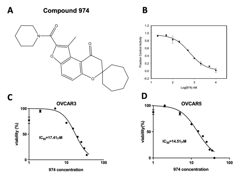
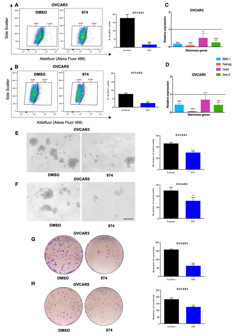
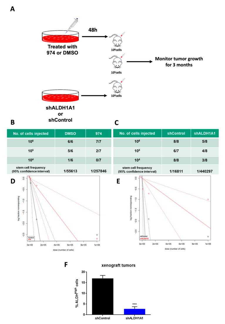
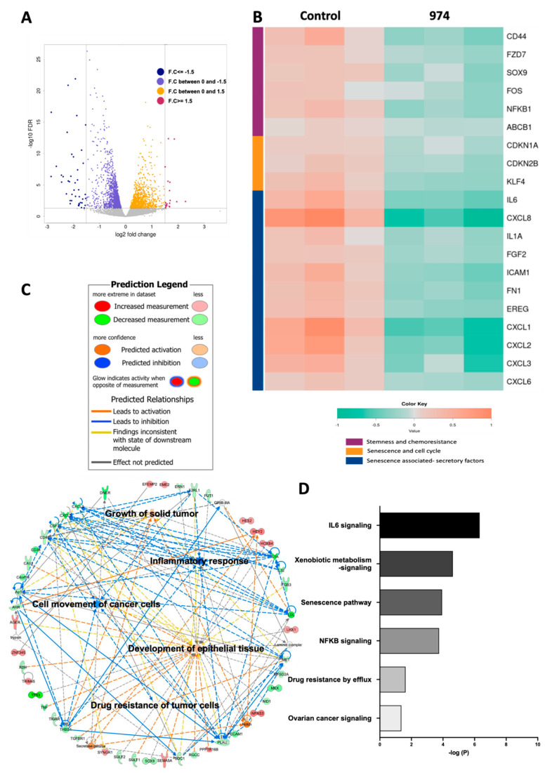
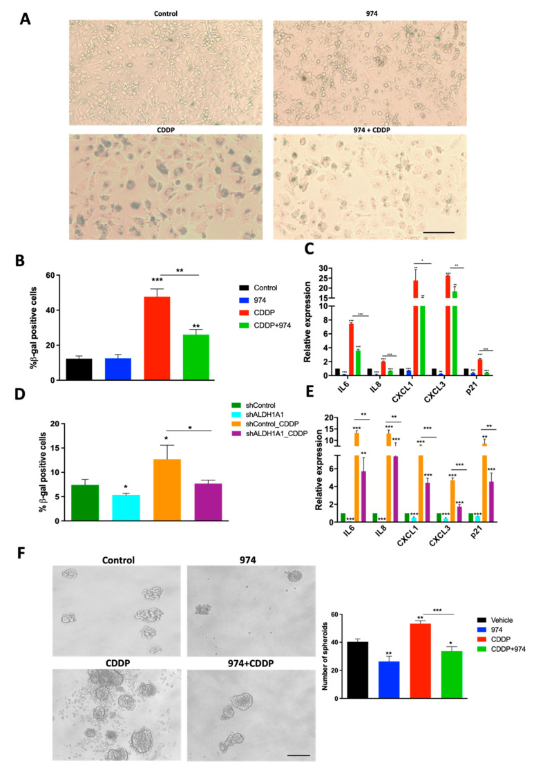
Ovarian cancer is a deadly disease attributed to late-stage detection as well as recurrence and the development of chemoresistance. Ovarian cancer stem cells (OCSCs) are hypothesized to be largely responsible for the emergence of chemoresistant tumors. Although chemotherapy may initially succeed at decreasing the size and number of tumors, it leaves behind residual malignant OCSCs. In this study, we demonstrate that aldehyde dehydrogenase 1A1 (ALDH1A1) is essential for the survival of OCSCs. We identified a first-in-class ALDH1A1 inhibitor, compound 974, and used 974 as a tool to decipher the mechanism of stemness regulation by ALDH1A1. The treatment of OCSCs with 974 significantly inhibited ALDH activity, the expression of stemness genes, and spheroid and colony formation. An in vivo limiting dilution assay demonstrated that 974 significantly inhibited CSC frequency. A transcriptomic sequencing of cells treated with 974 revealed a significant downregulation of genes related to stemness and chemoresistance as well as senescence and the senescence-associated secretory phenotype (SASP). We confirmed that 974 inhibited the senescence and stemness induced by platinum-based chemotherapy in functional assays. Overall, these data establish that ALDH1A1 is essential for OCSC survival and that ALDH1A1 inhibition suppresses chemotherapy-induced senescence and stemness. Targeting ALDH1A1 using small-molecule inhibitors in combination with chemotherapy therefore presents a promising strategy to prevent ovarian cancer recurrence and has the potential for clinical translation.

Keywords: ALDH1A1; cancer stem cells; chemotherapy resistance; ovarian cancer; senescence.


Conflict of interest statement

The authors declare no conflict of interest.

Figures

Figure 1
Figure 1
Compound 974: A novel ALDH1A1 inhibitor. (A) Chemical structure of compound 974. (B) EC50 curve for 974 binding with purified ALDH1A1. (C) OVCAR3 (D) OVCAR5 were treated with increasing doses of 974 (0.5–100 µM) for 48 h, and an MTT assay was performed to measure viability. IC50 values were calculated using GraphPad Prism.
Figure 2
Figure 2
ALDH1A1 inhibition suppresses ovarian cancer stemness phenotypes in vitro. (A) OVCAR3 or (B) OVCAR5 cells were treated with compound 974 (5 µM for 48 h) or DMSO. The percentage of ALDH+ cells was measured by an ALDEFLUOR assay using flow cytometry (left), and the results were quantified (right). (C) OVCAR3 or (D) OVCAR5 cells were treated as in A, and the expression of stemness genes was measured by qPCR. (E) OVCAR3 or (F) OVCAR5 cells were treated as in (A), and 500 cells/well were replated in 24-well low-adhesion conditions after treatment. Representative images of spheroid formation after 14 days (left) and quantification (right). (G) OVCAR3 or (H) OVCAR5 cells were treated as in (A), and 500 cells/well were replated in 6-well plates after treatment. Colonies were stained with 0.05% crystal violet and counted. Representative images of colony formation (left) and quantification (right). Error bars represent SEM; n = 3 independent experiments of triplicate assays. Data are presented as means ± SEM with p < 0.05 (*), p < 0.01 (**), and p < 0.005 (***). Scale bar, 100 µm.
Figure 3
Figure 3
ALDH1A1 inhibition suppresses ovarian cancer stemness in vivo. (A) Schematic representing study design. Injections of 106, 105, or 104 OVCAR3 cells treated with compound 974 (5 µM for 48 h), DMSO OR shALDH1A1, or shControl cells were given to NSG mice subcutaneously, and tumor formation was monitored. The numbers of mice with tumors over the total numbers of mice in the group and the CSC frequency were calculated by ELDA software (https://bioinf.wehi.edu.au/software/elda/, accessed on 12 May 2020) (B) for DMSO or 974 treatment and (C) for shControl or shALDH1A1. The log-fraction plot of limiting dilution analysis for stem cell frequency generated by extreme limiting dilution analysis in (D) compound 974 treatment vs DMSO or (E) shALDH1A1 or shControl. (F) The percentage of ALDH+ cells in the dissociated tumors from the shALDH1A1 study in (C) was measured by an ALDEFLUOR assay. Error bars represent SEM; n = 3 independent tumor samples. Data are presented as means ± SEM with p < 0.005 (***).
Figure 4
Figure 4
ALDH1A1 inhibition suppresses pathways involved in chemoresistance and stemness. RNA-seq was performed on OVCAR3 cells treated with compound 974 (5 µM for 48 h) or DMSO (n = 3). (A) Volcano plot of genes up and downregulated by ALDH1A1 inhibition. (B) Heatmap of selected genes significantly downregulated by compound 974 (FDR < 0.05). (C) Networks of biological processes constructed using significantly altered genes (FDR < 0.05) between OVCAR3 cells treated with 974 or DMSO. (D) Canonical pathways related to stemness and chemoresistance identified by the Ingenuity Pathway Analysis (IPA) using genes significantly altered by 974 treatment (FDR < 0.05, fold change > |1.5|).
Figure 5
Figure 5
ALDH1A1 inhibition suppresses chemotherapy-induced senescence and stemness in HGSOC cells. (A) Senescence-associated (SA) beta-gal assay was performed on OVCAR3 cells treated with DMSO, compound 974 (5 µM for 48 h), cisplatin (CDDP) (15 µM for 16 h), or both. Representative images at 10× magnification. (B) Quantification of SA-beta-gal assay represents percentage of senescent cells averaged from five different fields in each condition. (C) Expression of SASP genes and p21 (CIP1/WAF1) was examined by qPCR in OVCAR3 cells treated as in (A). (D) Percentage of SA-beta-gal-positive cells in shControl or shALDH1A1 cells treated with NaCl (vehicle) or CDDP (15 µM for 16 h) was measured by flow cytometry using Spider beta gal reagent. (E) Expression of p21 and SASP genes was examined by qPCR in shControl or shALDH1A1 cells treated with NaCl or CDDP (15 µM for 16h). (F) OVCAR3 cells treated with DMSO, compound 974 (5 µM for 48 h), cisplatin (CDDP) (7.5 µM; ½ IC50 for 3 h), or both were plated in low-attachment conditions at a density of 500 cells/well. Representative spheroid images captured on Day 7 (left). Quantification of spheroids (right). Error bars represent SEM; n = 3 independent experiments of triplicate assays. Data are presented as means ± SEM with p < 0.05 (*), p < 0.01 (**), and p < 0.005 (***). Scale bar, 100 µm.

References

    1. Torre L.A., Trabert B., DeSantis C.E., Miller K.D., Samimi G., Runowicz C.D., Gaudet M.M., Jemal A., Siegel R.L. Ovarian cancer statistics, 2018. CA Cancer J. Clin. 2018;68:284–296. doi: 10.3322/caac.21456. - DOI - PMC - PubMed
    1. Sung H., Ferlay J., Siegel R.L., Laversanne M., Soerjomataram I., Jemal A., Bray F. Global Cancer Statistics 2020: GLOBOCAN Estimates of Incidence and Mortality Worldwide for 36 Cancers in 185 Countries. CA Cancer J. Clin. 2021;71:209–249. doi: 10.3322/caac.21660. - DOI - PubMed
    1. Kurman R.J., Shih Ie M. The Dualistic Model of Ovarian Carcinogenesis: Revisited, Revised, and Expanded. Am. J. Pathol. 2016;186:733–747. doi: 10.1016/j.ajpath.2015.11.011. - DOI - PMC - PubMed
    1. Davis A., Tinker A.V., Friedlander M. “Platinum resistant” ovarian cancer: What is it, who to treat and how to measure benefit? Gynecol. Oncol. 2014;133:624–631. doi: 10.1016/j.ygyno.2014.02.038. - DOI - PubMed
    1. Steg A.D., Bevis K.S., Katre A.A., Ziebarth A., Dobbin Z.C., Alvarez R.D., Zhang K., Conner M., Landen C.N. Stem cell pathways contribute to clinical chemoresistance in ovarian cancer. Clin. Cancer Res. 2012;18:869–881. doi: 10.1158/1078-0432.CCR-11-2188. - DOI - PMC - PubMed