. 2022 Jul 1;12(7):876.
doi: 10.3390/brainsci12070876.

Escalation of Tau Accumulation after a Traumatic Brain Injury: Findings from Positron Emission Tomography

Affiliations

Escalation of Tau Accumulation after a Traumatic Brain Injury: Findings from Positron Emission Tomography

Abdalla Z Mohamed et al. Brain Sci. .

Abstract

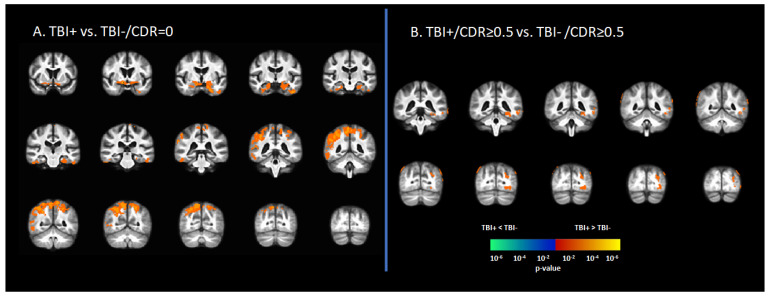
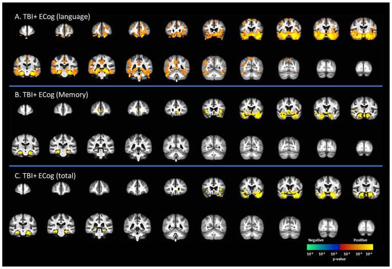
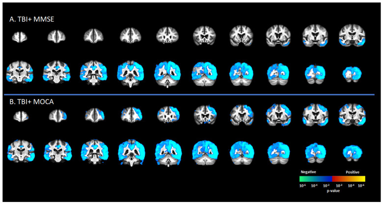
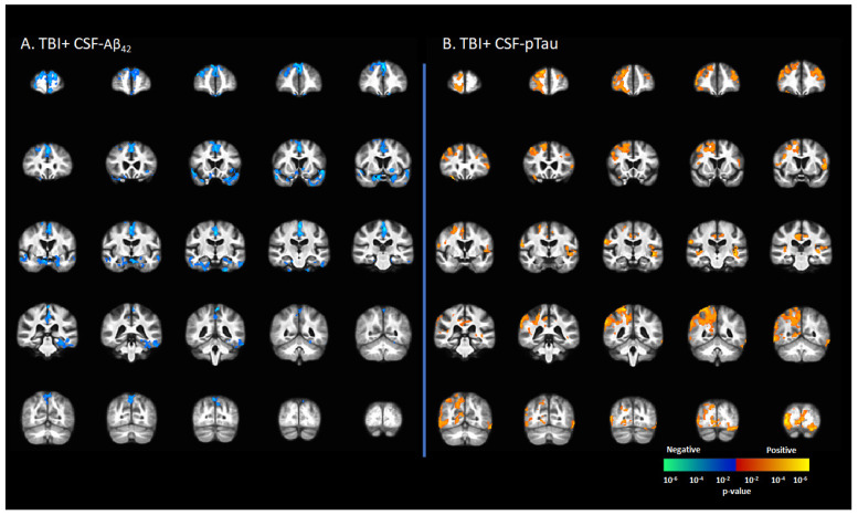
Traumatic brain injury (TBI) has come to be recognized as a risk factor for Alzheimer's disease (AD), with poorly understood underlying mechanisms. We hypothesized that a history of TBI would be associated with greater tau deposition in elders with high-risk for dementia. A Groups of 20 participants with self-reported history of TBI and 100 without any such history were scanned using [18F]-AV1451 positron emission tomography as part of the Alzheimer's Disease Neuroimaging Initiative (ADNI). Scans were stratified into four groups according to TBI history, and by clinical dementia rating scores into cognitively normal (CDR = 0) and those showing cognitive decline (CDR ≥ 0.5). We pursued voxel-based group comparison of [18F]-AV1451 uptake to identify the effect of TBI history on brain tau deposition, and for voxel-wise correlation analyses between [18F]-AV1451 uptake and different neuropsychological measures and cerebrospinal fluid (CSF) biomarkers. Compared to the TBI-/CDR ≥ 0.5 group, the TBI+/CDR ≥ 0.5 group showed increased tau deposition in the temporal pole, hippocampus, fusiform gyrus, and inferior and middle temporal gyri. Furthermore, the extent of tau deposition in the brain of those with TBI history positively correlated with the extent of cognitive decline, CSF-tau, and CSF-amyloid. This might suggest TBI to increase the risk for tauopathies and Alzheimer's disease later in life.

Keywords: Alzheimer’s disease; CFS-amyloid; CFS-tau; CSF biomarkers; cognitive decline; flortaucipir [18F]-AV1451; positron emission tomography; tau; traumatic brain injury.


Conflict of interest statement

The authors declare no conflict of interest.

Figures

Figure 1
Figure 1
Differences in tau deposition to [18F]-AV1451 PET between participants with self-reported history of traumatic brain injury (TBI+) and those without TBI history (TBI-). (A) The statistical difference map of [18F]-AV1451 SUVr between the TBI+ group in contrast to the control group (TBI-/CDR = 0) group, showing that history of TBI increased the tau deposition in wide-spread brain regions. (B) The statistical difference map of [18F]-AV1451 SUVr in the symptomatic cases between the TBI+/CDR ≥ 0.5 (in contrast to TBI-/CDR ≥ 0.5) showed increased tau deposition in cortical regions overlapping with those reported for Alzheimer’s disease [40,41]. The statistical tests were corrected for nuisance covariates, including age, gender, education, and APOE-ε4 status. Red-yellow shows regions with the TBI+ group showing higher tau as compared to TBI-, while blue-green represents less tau deposition in the TBI+ as compared to TBI-.
Figure 2
Figure 2
Correlation between [18F]-AV1451 SUVr maps and everyday cognition (ECog) scores in the population with a self-reported history of traumatic brain injury (TBI), with results of the correlations between tau deposition and (A) ECog-Language sub-test, (B) ECog-Memory sub-test, and (C) ECog-total score. Red-yellow represents a positive correlation between tau accumulation in people with self-reported history of TBI and ECog scores, while blue-green represents negative correlation between tau accumulation and ECog score.
Figure 3
Figure 3
Correlation between [18F]-AV1451 SUVr maps and different cognitive test scores in the elderly population with a self-reported history of traumatic brain injury (TBI), with results showing negative correlations between tau deposition and (A) mini-mental state scale (MMSE) and (B) Montreal cognitive assessment (MOCA). Red-yellow represents a positive correlation between tau accumulation in people with self-reported history of TBI and cognitive scores, while blue-green represents negative correlation between tau accumulation and cognitive scores.
Figure 4
Figure 4
Correlation between [18F]-AV1451 SUVr maps and cerebrospinal fluid (CSF) biomarkers in the population with a self-reported history of traumatic brain injury (TBI), with results showing (A) negative correlations between CSF-Aβ42 and [18F]-AV1451 SUVr in brain; (B) positive correlation between CSF-pTau and [18F]-AV1451 SUVr in the brain. Red-yellow represents a positive correlation between tau accumulation in TBI+ cases and CSF biomarkers, while blue-green represents negative correlation between tau accumulation and CSF biomarkers.

References

    1. Shen Q., Hiebert J.B., Hartwell J., Thimmesch A.R., Pierce J.D. Systematic Review of Traumatic Brain Injury and the Impact of Antioxidant Therapy on Clinical Outcomes. Worldviews Evid. -Based Nurs. 2016;13:380–389. doi: 10.1111/wvn.12167. - DOI - PubMed
    1. Mohamed A.Z., Corrigan F., Collins-Praino L.E., Plummer S.L., Soni N., Nasrallah F.A. Evaluating Spatiotemporal Microstructural Alterations Following Diffuse Traumatic Brain Injury. NeuroImage Clin. 2020;25:102136. doi: 10.1016/j.nicl.2019.102136. - DOI - PMC - PubMed
    1. Mohamed A.Z., Cumming P., Nasrallah F.A. White Matter Alterations Are Associated With Cognitive Dysfunction Decades After Moderate-to-Severe Traumatic Brain Injury and/or Posttraumatic Stress Disorder. Biol. Psychiatry Cogn. Neurosci. Neuroimaging. 2021;6:1100–1109. doi: 10.1016/j.bpsc.2021.04.014. - DOI - PubMed
    1. Mohamed A.Z., Cumming P., Nasrallah F.A. Traumatic Brain Injury Augurs Ill for Prolonged Deficits in the Brain’s Structural and Functional Integrity Following Controlled Cortical Impact Injury. Sci. Rep. 2021;11:1–13. doi: 10.1038/s41598-021-00660-5. - DOI - PMC - PubMed
    1. To X.V., Mohamed A.Z., Cumming P., Nasrallah F.A. Subacute Cytokine Changes after a Traumatic Brain Injury Predict Chronic Brain Microstructural Alterations on Advanced Diffusion Imaging in the Male Rat. Brain. Behav. Immun. 2022 doi: 10.1016/j.bbi.2022.02.017. - DOI - PubMed