. 2022 Jul 13;12(7):920.
doi: 10.3390/brainsci12070920.

Three-Hour Argon Inhalation Has No Neuroprotective Effect after Open Traumatic Brain Injury in Rats

Affiliations

Three-Hour Argon Inhalation Has No Neuroprotective Effect after Open Traumatic Brain Injury in Rats

Viktoriya V Antonova et al. Brain Sci. .

Abstract

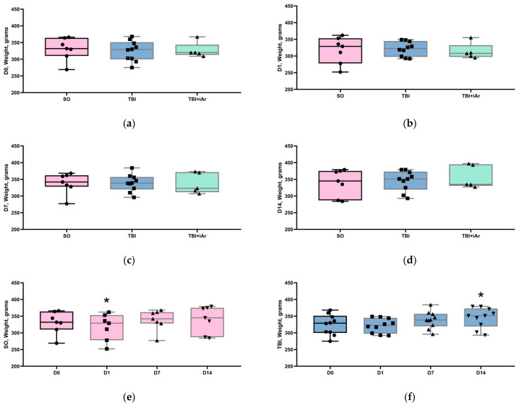
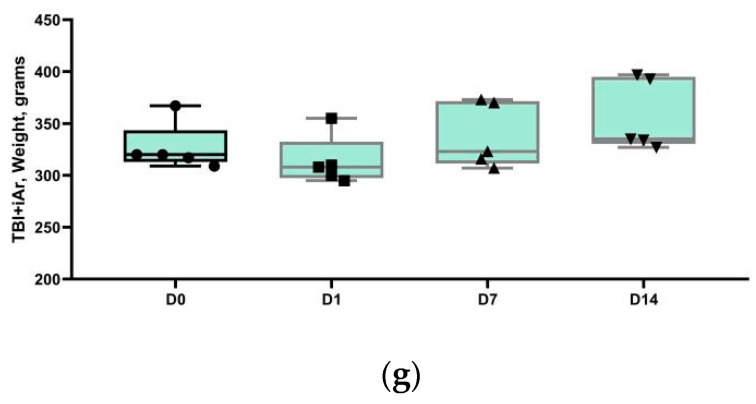
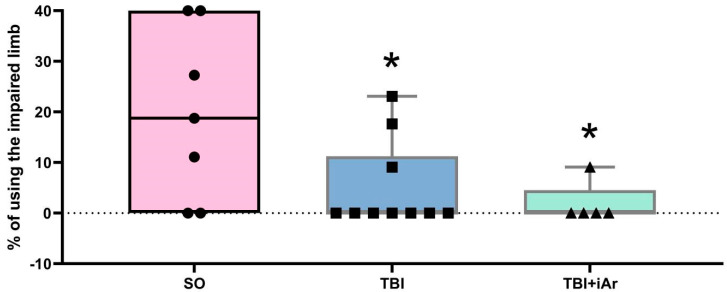
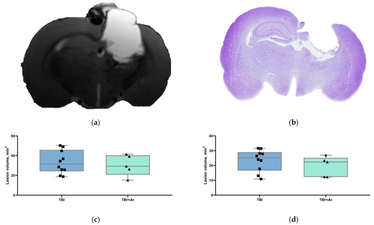
In vivo studies of the therapeutic effects of argon in traumatic brain injury (TBI) are limited, and their results are contradictory. The aim of this study was to evaluate the effect of a three-hour inhalation of argon (70%Ar/30%O2) after an open TBI on the severity of the neurological deficit and the degree of brain damage in rats. The experiments were performed on male Wistar rats (n = 35). The TBI was simulated by the dosed open brain contusion injury. The animals were divided into three groups: sham-operated (SO, n = 7); TBI + 70%N2/30%O2 (TBI, n = 14); TBI + 70%Ar/30%O2 (TBI + iAr, n = 14). The Neurological status was assessed over a 14-day period (using the limb-placing and cylinder tests). Magnetic resonance imaging (MRI) scans and a histological examination of the brain with an assessment of the volume of the lesions were performed 14 days after the injury. At each of the time points (days 1, 7, and 14), the limb-placing test score was lower in the TBI and TBI + iAr groups than in the SO group, while there were no significant differences between the TBI and TBI + iAr groups. Additionally, no differences were found between these groups in the cylinder test scores (day 13). The volume of brain damage (tissue loss) according to both the MRI and histological findings did not differ between the TBI and TBI + iAr groups. A three-hour inhalation of argon (70%Ar/30%O2) after a TBI had no neuroprotective effect.

Keywords: argon; neuroprotection; organoprotection; traumatic brain injury.


Conflict of interest statement

The authors declare no conflict of interest.

Figures

Figure 1
Figure 1
Analysis of body weight data. (a) Weight of rats the day before the simulated TBI; (b) weight of rats on the day of the simulated TBI; (c) weight of rats 7 days after the simulated TBI; (d) weight of rats 14 days after the simulated TBI; (e) weight change in the control group; (f) weight change in the TBI group; (g) weight change in the TBI + iAr group. The data are presented as median, quartiles, and single values. *—p < 0.05 vs. D0, Kruskal–Wallis test with Dunn’s multiple comparisons. ■; ●; ▲; ▼—single values within specific statistical sample.
Figure 1
Figure 1
Analysis of body weight data. (a) Weight of rats the day before the simulated TBI; (b) weight of rats on the day of the simulated TBI; (c) weight of rats 7 days after the simulated TBI; (d) weight of rats 14 days after the simulated TBI; (e) weight change in the control group; (f) weight change in the TBI group; (g) weight change in the TBI + iAr group. The data are presented as median, quartiles, and single values. *—p < 0.05 vs. D0, Kruskal–Wallis test with Dunn’s multiple comparisons. ■; ●; ▲; ▼—single values within specific statistical sample.
Figure 2
Figure 2
(a) LPT score 1 day after the simulation of TBI; (b) LPT score 7 days after the simulation of TBI; (c) LPT score 14 days after the simulation of TBI; (d) changes in LPT score in the control group; (e) changes in LPT score in the TBI group; (f) changes in LPT score in the TBI + iAr group. The data are presented as median, quartiles, and single values. *—p < 0.05 vs. SO group on (ac) figures, vs. D1 group on (df) figures; #—p < 0.05 vs. D7 group, Kruskal–Wallis test with Dunn’s multiple comparisons. ■; ●; ▲—single values within specific statistical sample.
Figure 2
Figure 2
(a) LPT score 1 day after the simulation of TBI; (b) LPT score 7 days after the simulation of TBI; (c) LPT score 14 days after the simulation of TBI; (d) changes in LPT score in the control group; (e) changes in LPT score in the TBI group; (f) changes in LPT score in the TBI + iAr group. The data are presented as median, quartiles, and single values. *—p < 0.05 vs. SO group on (ac) figures, vs. D1 group on (df) figures; #—p < 0.05 vs. D7 group, Kruskal–Wallis test with Dunn’s multiple comparisons. ■; ●; ▲—single values within specific statistical sample.
Figure 3
Figure 3
The cylinder test results. Day 13 day after the traumatic brain injury. The data are presented as median, quartiles, and single values. *—p < 0.05 vs. SO group, Mann–Whitney U-test with Bonferroni correction. ■; ●; ▲—single values within specific statistical sample.
Figure 4
Figure 4
(a) Representative T2-weighted coronal MR-images and (b) cresyl violet-stained sections of the rat brain on day 14 post-TBI demonstrating loss of cortical tissue at the site of impact. (c) Volume of brain lesions caused by traumatic brain injury in the TBI and TBI + iAr groups based on MRI data. (d) Volume of brain lesions caused by traumatic brain injury in the TBI and TBI + iAr groups based on histological examination data. No significant difference between the groups was found (Mann–Whitney U criterion). The data are presented as median, quartiles, and single values. ■; ▲—single values within specific statistical sample.

References

    1. Maas A.I.R., Menon D.K., Adelson P.D., Andelic N., Bell M.J., Belli A., Bragge P., Brazinova A., Büki A., Chesnut R.M., et al. Traumatic brain injury: Integrated approaches to improve prevention, clinical care, and research. Lancet Neurol. 2017;16:987–1048. doi: 10.1016/S1474-4422(17)30371-X. - DOI - PubMed
    1. Talypov A.E., Grin’ A.A., Petrikov S.S., Krylov V.V., Solodov A.A., Kordonsky A.Ю., Shabanov A.K., Barmina T.G., Mullagulov T.R. Intracranial pressure monitoring in patients with severe head injury. Russ. J. Neurosurg. 2021;22:14–27. doi: 10.17650/1683-3295-2020-22-4-14-27. - DOI
    1. Ovsyannikov D.M., Chekhonatsky A.A., Kolesov V.N., Bubashvili A.I. Social and Epidemiological Aspects of Craniocerebral Trauma (review) Saratov J. Med. Sci. Res. 2012;8:777–785. (In Russian)
    1. Ng S.Y., Lee A.Y.W. Traumatic Brain Injuries: Pathophysiology and Potential Therapeutic Targets. Front. Cell. Neurosci. 2019;13:528. doi: 10.3389/fncel.2019.00528. - DOI - PMC - PubMed
    1. Liu J., Nolte K., Brook G., Liebenstund L., Weinandy A., Höllig A., Veldeman M., Willuweit A., Langen K.-J., Rossaint R., et al. Post-stroke treatment with argon attenuated brain injury, reduced brain inflammation and enhanced M2 microglia/macrophage polarization: A randomized controlled animal study. Crit. Care. 2019;23:198. doi: 10.1186/s13054-019-2493-7. - DOI - PMC - PubMed