Review
. 2022 Jul 15;10(7):1709.
doi: 10.3390/biomedicines10071709.

Mutation of Proteolipid Protein 1 Gene: From Severe Hypomyelinating Leukodystrophy to Inherited Spastic Paraplegia

Affiliations
Review

Mutation of Proteolipid Protein 1 Gene: From Severe Hypomyelinating Leukodystrophy to Inherited Spastic Paraplegia

Guy Khalaf et al. Biomedicines. .

Abstract

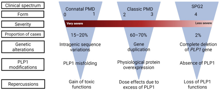
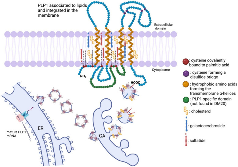
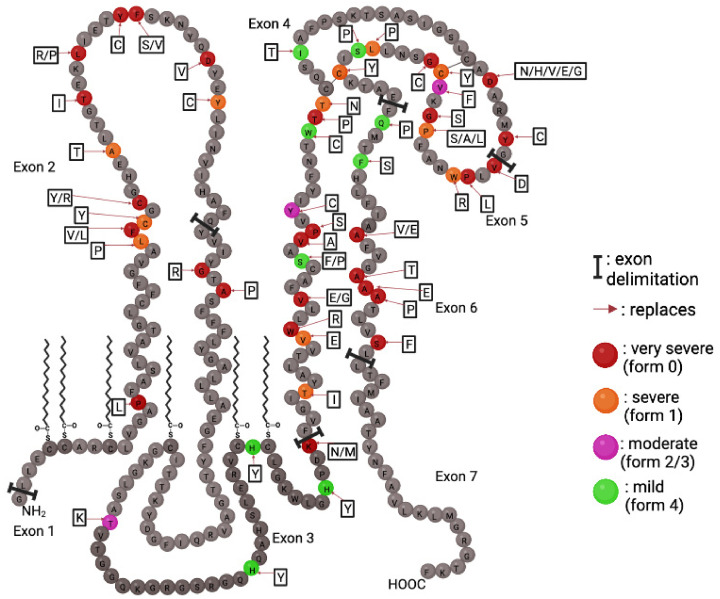
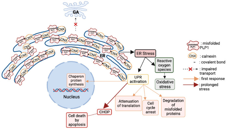
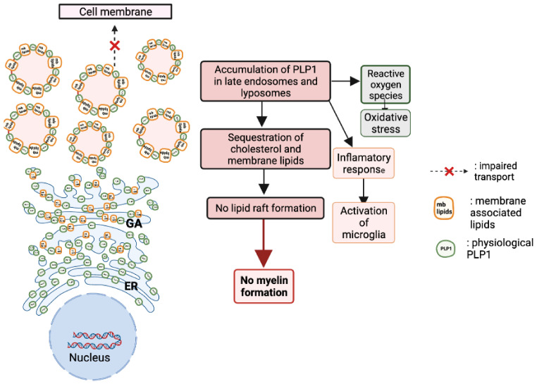
Pelizaeus-Merzbacher Disease (PMD) is an inherited leukodystrophy affecting the central nervous system (CNS)-a rare disorder that especially concerns males. Its estimated prevalence is 1.45-1.9 per 100,000 individuals in the general population. Patients affected by PMD exhibit a drastic reduction or absence of myelin sheaths in the white matter areas of the CNS. The Proteolipid Protein 1 (PLP1) gene encodes a transmembrane proteolipid protein. PLP1 is the major protein of myelin, and it plays a key role in the compaction, stabilization, and maintenance of myelin sheaths. Its function is predominant in oligodendrocyte development and axonal survival. Mutations in the PLP1 gene cause the development of a wide continuum spectrum of leukopathies from the most severe form of PMD for whom patients exhibit severe CNS hypomyelination to the relatively mild late-onset type 2 spastic paraplegia, leading to the concept of PLP1-related disorders. The genetic diversity and the biochemical complexity, along with other aspects of PMD, are discussed to reveal the obstacles that hinder the development of treatments. This review aims to provide a clinical and mechanistic overview of this spectrum of rare diseases.

Keywords: Pelizaeus-Merzbacher disease (PMD); animal models; diagnosis; proteolipid protein 1 variants; spastic paraplegia (SPG2); treatments.


Conflict of interest statement

The authors declare no conflict of interest.

Figures

Figure 1
Figure 1
Clinical classification of PLP1-related disorders. The spectrum of the disease is a continuum ranging from severe PMD (form 0) to SPG2 (form 4), with decreasing clinical severity. Under each category, the most frequent PLP1 mutations and their consequences are indicated.
Figure 2
Figure 2
MRI of a 5-year-old Pelizaeus–Merzbacher Disease (PMD) patient in comparison with an age-matched normal boy (Normal). In physiological conditions, the myelinated white matter has a hypersignal T1, hyposignal T2, and flair when compared to gray matter. In PMD, the hypomyelinated white matter has a normal hypersignal T1 appearance contrasting with an abnormal hypersignal T2 and flair. The corpus callosum is partially myelinated with a hyposignal T2 but not with flair.
Figure 3
Figure 3
The physiological state of PLP1 formation: from mRNA to a functional transmembrane protein. The mature mRNA encoding PLP1 is translated in the rough endoplasmic reticulum (ER). Afterward, the immature protein is transported to the Golgi apparatus (GA), where it will associate with lipidic myelin components, such as cholesterol, galactocerebrosides, and sulfatides to form lipidic rafts. Then, the lipidic rafts are sent toward the cellular membrane to form the myelin sheath. PLP1 is a 30 kDa tetraspan transmembrane protein with NH2 and COOH termini in the cytoplasm. It has four transmembrane α-helices inside the membrane of oligodendrocytes, and it is a highly hydrophobic protein. Palmitic acid is covalently bound to PLP1 via six cysteine residues by an autocatalytic posttranslational modification. The fatty acids attached to the intracellular loop of PLP1 ensure the integration of this protein in the lipid leaflet in compact myelin.
Figure 4
Figure 4
Point mutations of PLP1 and the severity of their repercussions. PLP1 point mutations are color coded to reflect the severity of the caused pathological phenotype. Mutations can occur throughout all seven exons. Amino acid changes are shown in adjacent boxes.
Figure 5
Figure 5
Consequences of PLP1 point mutations on the cellular physiology. The mutant PLP1 accumulates inside the oligodendrocytes in the endoplasmic reticulum (ER), and they cannot be delivered to the cell membrane. The accumulation is caused by the stable biding of a PLP1 transmembrane domain with calnexin, an ER chaperon protein. The prolonged binding of these two proteins prevents the proper functioning of the ER-associated protein degradation pathway to correctly eliminate the misfolded proteins. Moreover, the cytoplasmic accumulation of misfolded PLP1 and DM20 activates the unfolded protein response (UPR) pathway that aims to rescue the cell by stopping protein translation, degrading misfolded proteins, and increasing the production of chaperon proteins responsible of protein folding. When all the rescue mechanisms of the UPR are in vain, the ultimate response is to induce apoptosis by the activation of the CHOP pathway.
Figure 6
Figure 6
Consequences of PLP1 duplication on the cellular physiology. When overexpressed, PLP1 sequesters cholesterol and accumulates in late endosomes and lysosomes. Moreover, raft formation and membrane trafficking are crucial for myelin formation, and their disruption causes the loss of myelin. In addition to its cytotoxic effects, PLP1 protein overexpression leads to an inflammatory response with the activation of microglia in white and grey matter and the upregulation of cytokines and their receptors. It is probable that the microglial activation and the inflammatory response contribute to a certain extent to the pathology’s development and progression.

References

    1. Raine C.S. Myelin. Springer; Berlin/Heidelberg, Germany: 1984. Morphology of myelin and myelination; pp. 1–50.
    1. Lemke G. Unwrapping the genes of myelin. Neuron. 1988;1:535–543. doi: 10.1016/0896-6273(88)90103-1. - DOI - PubMed
    1. Nave K.A. Myelination and the trophic support of long axons. Nat. Rev. Neurosci. 2010;11:275–283. doi: 10.1038/nrn2797. - DOI - PubMed
    1. Lappe-Siefke C., Goebbels S., Gravel M., Nicksch E., Lee J., Braun P.E., Griffiths I.R., Nave K.-A. Disruption of Cnp1 uncouples oligodendroglial functions in axonal support and myelination. Nat. Genet. 2003;33:366–374. doi: 10.1038/ng1095. - DOI - PubMed
    1. van der Knaap M.S., Bugiani M. Leukodystrophies: A proposed classification system based on pathological changes and pathogenetic mechanisms. Acta Neuropathol. 2017;134:351–382. doi: 10.1007/s00401-017-1739-1. - DOI - PMC - PubMed