Review
. 2022 Jun 29;12(7):1581.
doi: 10.3390/diagnostics12071581.

Myelodysplastic Syndrome: Diagnosis and Screening

Affiliations
Review

Myelodysplastic Syndrome: Diagnosis and Screening

Francisco P Tria 4th et al. Diagnostics (Basel). .

Abstract

Myelodysplastic syndromes (MDS) are heterogeneous groups of clonal myeloid disorders characterized by unexplained persistent peripheral blood (PB) cytopenia(s) of one or more of the hematopoietic lineages, or bone marrow (BM) morphologic dysplasia in hematopoietic cells, recurrent genetic abnormalities, and an increased risk of progression to acute myeloid leukemia (AML). In the past several years, diagnostic, prognostic, and therapeutic approaches have substantially improved with the development of Next Generation Sequencing (NGS) diagnostic testing and new medications. However, there is no single diagnostic parameter specific for MDS, and correlations with clinical information, and laboratory test findings are needed to reach the diagnosis.

Keywords: cytogenetics; myelodysplastic syndromes; next generation sequencing.


Conflict of interest statement

The authors declare no conflict of interest.

Figures
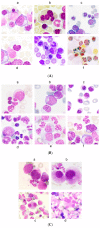
Figure 1
Figure 1
(A) Erythroid dysplasia (100×). (a) megaloblastosis; (b) multinuclearity; (c) nuclear lobulation; (d) pyknosis; (e) defective hemoglobinization and cytoplasmic fraying; (f) ring sideroblasts. Adapted from Della Porta et al. [9]; (B) Granulocytic dysplasia (100×). (a) myeloblast; (b) Auer rod; (c) hypolobulation; (d,e) abnormal nuclear shape; (f) hypogranulation. Adapted from Della Porta et al. [9]; (C) Megakaryocytic dysplasia (100×). (a) micromegakaryocyte; (b) monolobated megakaryocyte; (c) small binucleated megakaryocyte; (d) megakaryocyte with multiple separated nuclei. Adapted from Della Porta et al. [9].
Figure 2
Figure 2
(A) Shows normal maturational pattern of nucleated hematopoietic cellular populations. Regenerating myeloblasts showing intact CD34 and CD117 expression and heterogeneous CD33 positivity. Maturing granulocytic precursors showing the classic “Nike swoosh” pattern in the CD13/CD16 plot, and monocytes showing intact expression of CD14, without aberrant antigen expression. Lymphocytes in green, monocytes in orange, granulocytes in blue, blasts in red; (B) Case of MDS showing myeloblasts with loss of CD117 and with concurrently bright CD33 intensity. Prominent abnormal granulocytic maturational pattern in CD13/CD16 plot, losing the classic “Nike swoosh” pattern. In this MDS case, the granulocytes and monocytes also show aberrant CD56 expression; Lymphocytes in green, monocytes in orange, granulocytes in blue, blasts in red.
Figure 2
Figure 2
(A) Shows normal maturational pattern of nucleated hematopoietic cellular populations. Regenerating myeloblasts showing intact CD34 and CD117 expression and heterogeneous CD33 positivity. Maturing granulocytic precursors showing the classic “Nike swoosh” pattern in the CD13/CD16 plot, and monocytes showing intact expression of CD14, without aberrant antigen expression. Lymphocytes in green, monocytes in orange, granulocytes in blue, blasts in red; (B) Case of MDS showing myeloblasts with loss of CD117 and with concurrently bright CD33 intensity. Prominent abnormal granulocytic maturational pattern in CD13/CD16 plot, losing the classic “Nike swoosh” pattern. In this MDS case, the granulocytes and monocytes also show aberrant CD56 expression; Lymphocytes in green, monocytes in orange, granulocytes in blue, blasts in red.
Figure 3
Figure 3
(A) CHIP as a precursor to hematologic neoplasms. Sequential acquisition of somatic mutations leading to clonal instability and survival advantage of mutated cells leading to expansion of neoplastic clones. Adapted from Steensma et al. [85]; (B) Cumulative incidence of hematologic malignancies. With increasing number of somatic mutations acquired during aging (CHIP), there is a proportional increase in the risk of developing hematologic malignancies. Adapted from Jaiswal et al. [80].
Figure 4
Figure 4
Correlation of number of co-mutations in cytogenetic prognostic-stratified MDS. Two or more mutations identified in 69% of cytogenetically normal MDS (blue), occurring in up to 11 mutant genes in a single patient. Adapted from Tria et al. [58].
Figure 5
Figure 5
(A,B). Data showing AML transformation occurring significantly faster in patients who acquire histone modifier gene and signaling gene mutations at initial diagnosis. Adapted from Tria et al. [58].
Figure 6
Figure 6
Kaplan–Meier curves in IPSS-M showing significant differences in probability of leukemia-free survival in each subgroup. Adapted from Bernard et al. [99].
Figure 7
Figure 7
Detection of dysplasia by AI. (100×) (A) The detectors distinguish the cells of interest (green boxes include all nucleated cells and red boxes include red blood cells, platelets, and debris); (B) Normal mature neutrophil with adequate cytoplasmic granules (left) and granulocytic hypogranularity in a mature neutrophil (right). Adapted from Mori et al. [103].

References

    1. Olcay L., Yetgin S. Disorders Mimicking Myelodysplastic Syndrome and Difficulties in Its Diagnosis. IntechOpen; London, UK: 2016. - DOI
    1. Steensma D.P. Dysplasia has A differential diagnosis: Distinguishing genuine myelodysplastic syndromes (MDS) from mimics, imitators, copycats and impostors. Curr. Hematol. Malig. Rep. 2012;7:310–320. doi: 10.1007/s11899-012-0140-3. - DOI - PubMed
    1. Arber D.A., Orazi A., Hasserjian R., Thiele J., Borowitz M.J., Le Beau M.M., Bloomfield C.D., Cazzola M., Vardiman J.W. The 2016 revision to the World Health Organization classification of myeloid neoplasms and acute leukemia. Blood. 2016;127:2391–2405. doi: 10.1182/blood-2016-03-643544. - DOI - PubMed
    1. Greenberg P., Cox C., LeBeau M.M., Fenaux P., Morel P., Sanz G., Sanz M., Vallespi T., Hamblin T., Oscier D., et al. International scoring system for evaluating prognosis in myelodysplastic syndromes. Blood. 1997;89:2079–2088. doi: 10.1182/blood.V89.6.2079. - DOI - PubMed
    1. Greenberg P.L., Tuechler H., Schanz J., Sanz G., Garcia-Manero G., Sole F., Bennett J.M., Bowen D., Fenaux P., Dreyfus F., et al. Revised international prognostic scoring system for myelodysplastic syndromes. Blood. 2012;120:2454–2465. doi: 10.1182/blood-2012-03-420489. - DOI - PMC - PubMed