. 2022 Jul 7;12(7):1653.
doi: 10.3390/diagnostics12071653.

Do Not Miss Acute Diffuse Panbronchiolitis for Tree-in-Bud: Case Series of a Rare Lung Disease

Affiliations

Do Not Miss Acute Diffuse Panbronchiolitis for Tree-in-Bud: Case Series of a Rare Lung Disease

Johannes Raedler et al. Diagnostics (Basel). .

Abstract

Acute bronchiolitis is a common disease of infants affecting the small airways. Rarely, acute bronchiolitis may occur in adolescents and adults. Here, we present four unrelated adolescent patients with severe clinical presentation and unique CT imaging with extensive tree-in-bud pattern, representing a rare clinical phenotype of acute diffuse panbronchiolitis. This characteristic disease pattern caused by inhalation injury from waterpipes, smoked tobacco, and cannabinoids must be differentiated from e-cigarette or vaping product-use-associated lung injury (EVALI). Visual diagnosis of CT and an early diagnostic procedure for detection and differentiation of inhaled hazards, including sample storage for future identification of novel noxious agents, are warranted.

Keywords: acute bronchiolitis; acute diffuse panbronchiolitis; case report; pediatric pulmonology; pulmonology; respiratory failure; tree-in-bud pattern.


Conflict of interest statement

The authors declare no conflict of interest.

Figures

Figure 1
Figure 1
Clinical courses and spirometry results. (A) Results of pCO2 from blood gas analysis and C-reactive protein (CRP) from laboratory studies during hospitalization. Antibiotic treatment intervals are indicated. Additionally, the patients received systemic prednisolone for reduction in airway inflammation. All patients showed a normalization of pCO2 and CRP levels prior to discharge. (B) Spirometry results during follow-up and methylprednisolone burst treatments for Patients 1 and 2 (dosage indicated, each as daily dosage for 3 days). Initial spirometry showed peripheral obstruction, overinflation, increased airway resistance, and pseudorestriction in both patients. No spirometry follow-up was available for Patient 3 or 4. amp = ampicillin, ama = ampicillin/sulbactam, azm = azithromacin, cef = cefpodoxime, clr = clarithromycin, dox = doxycycline, CT = computed tomography, mem = meropenem, mpr = methylprednisolone burst, mxf = moxifloxacin, ptz = piperacillin-tazobactam, pred = prednisolone, and van = vancomycin.
Figure 2
Figure 2
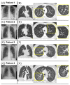
Radiographic findings. (A,B) Radiographic findings for Patient 1. Chest X-ray on day 2 showing signs of peribronchial cuffing (A). Coronal CT image on day 8 shows diffuse and homogenous distribution of centrilobular micronodules and tree-in-bud pattern through the whole lung (B, left). Axial maximal intensity projection (MIP, 5 mm thickness) of the CT scan improves the detection and distribution of the disease pattern (B, right). In the magnified view, the tree-in-bud pattern is mostly explained by peribronchial thickening. (C,D) Radiographic findings for Patient 2. Chest X-ray on day 1 shows slight enlargement of the hilar lymph nodes, increased peribronchial and interstitial markings, small opacification in the right basal area, and small bilateral pleural effusions (F). Coronal CT image on day 4 (G, left) and axial MIP magnification (G, right) show diffuse and homogenous tree-in-bud pattern, mostly due to endobronchial pathology (G). (E,F) Radiographic findings for Patient 3. Chest X-ray on day 6 shows diffuse bilateral peribronchial markings with basal dominance and small pleural effusions suspicious for atypical pneumonia (E). Coronal CT image on day 6 (F, left) and axial MIP magnification (F, right) show tree-in-bud pattern due to peribronchial thickening with diffuse and homogenous distribution through the whole lung. (G,H) Radiographic findings for Patient 4 (provided by the Department of Radiology, Medical University Innsbruck, Austria). Chest X-ray on day 1 shows bilateral widespread nodular opacities (G). Coronal CT image on day 2 (H, left) and axial MIP magnification (H, right) show diffuse and homogenous tree-in-bud pattern through the whole lung caused by peribronchial thickening (H).
Figure 3
Figure 3
CT follow-up. (A) Follow-up CT of Patient 1 after 6 months showed near complete resolution of the previously extensive tree-in-bud pattern. (B) Follow-up CT of Patient 2 after 35 months showed no tree-in-bud pattern but centrilobular nodular hazy densities with upper lobe predominance suspicious for respiratory bronchiolitis disease, possibly secondary to inhaled noxae. (C) Follow-up CT of Patient 3 after two months showed significant improvement. Due to the recent manifestation, no follow-up CT of Patient 4 was currently available.

Similar articles

References

    1. Florin T.A., Plint A.C., Zorc J.J. Viral bronchiolitis. Lancet. 2017;389:211–224. doi: 10.1016/S0140-6736(16)30951-5. - DOI - PMC - PubMed
    1. Ryu J.H., Myers J.L., Swensen S.J. Bronchiolar disorders. Am. J. Respir. Crit. Care Med. 2003;168:1277–1292. doi: 10.1164/rccm.200301-053SO. - DOI - PubMed
    1. Winningham P.J., Martínez-Jiménez S., Rosado-de-Christenson M.L., Betancourt S.L., Restrepo C.S., Eraso A. Bronchiolitis: A Practical Approach for the General Radiologist. Radiographics. 2017;37:777–794. doi: 10.1148/rg.2017160131. - DOI - PubMed
    1. Poletti V., Chilosi M., Casoni G., Colby T.V. Diffuse panbronchiolitis. Sarcoidosis Vasc Diffus. Lung Dis. 2004;21:94–104. doi: 10.1183/09031936.06.00131805. - DOI - PubMed
    1. Poletti V., Casoni G., Chilosi M., Zompatori M. Diffuse panbronchiolitis. Eur. Respir. J. 2006;28:862–871. doi: 10.1183/09031936.06.00131805. - DOI - PubMed

LinkOut - more resources