

Diagnostic Accuracy of PET/CT or PET/MRI Using PSMA-Targeting Radiopharmaceuticals in High-Grade Gliomas: A Systematic Review and a Bivariate Meta-Analysis
- PMID: 35885569
- PMCID: PMC9323081
- DOI: 10.3390/diagnostics12071665
Diagnostic Accuracy of PET/CT or PET/MRI Using PSMA-Targeting Radiopharmaceuticals in High-Grade Gliomas: A Systematic Review and a Bivariate Meta-Analysis
Abstract
Background: Several studies proposed the use of positron emission tomography (PET) with Prostate Specific Membrane Antigen (PSMA)-targeting radiopharmaceuticals in brain tumors. Our aim is to calculate the diagnostic accuracy of these methods in high-grade gliomas (HGG) with a bivariate meta-analysis.
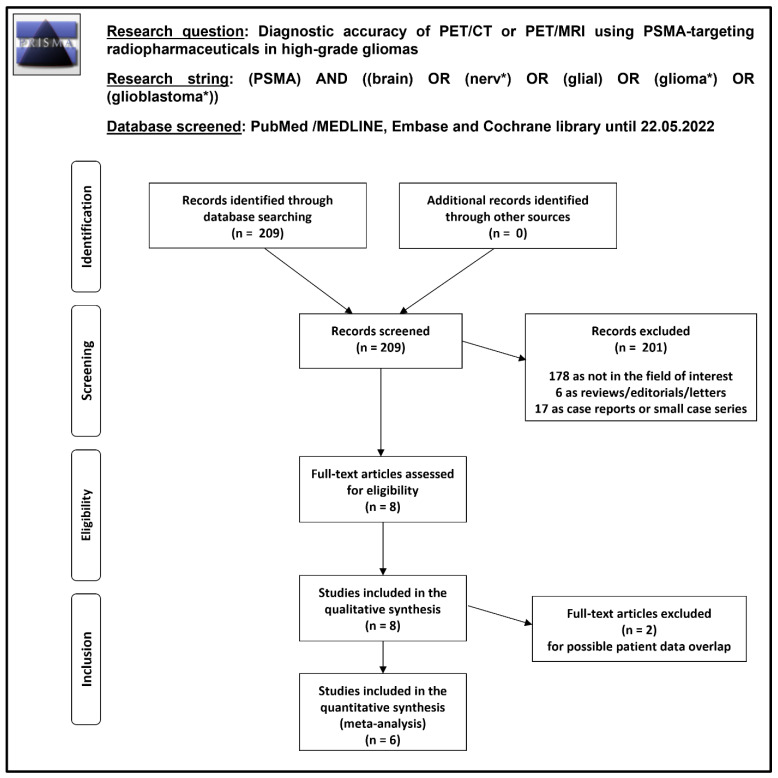
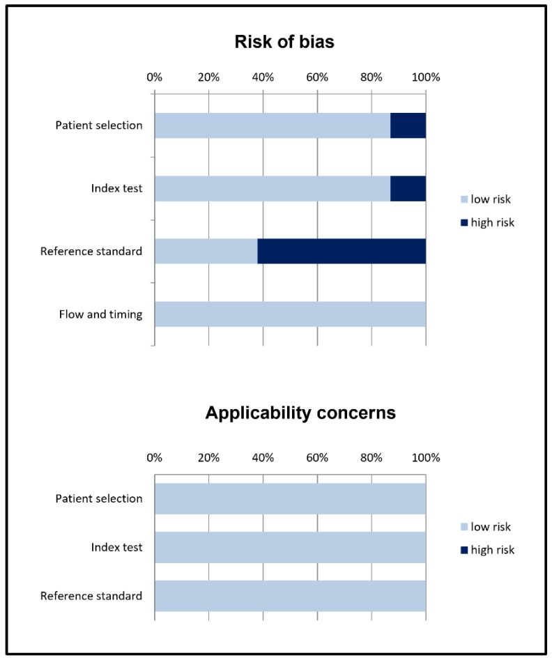
Methods: A comprehensive literature search of studies on the diagnostic accuracy of PET/CT or PET/MRI with PSMA-targeting radiopharmaceuticals in HGG was performed. Original articles evaluating these imaging methods both in the differential diagnosis between HGG and low-grade gliomas (LGG) and in the assessment of suspicious HGG recurrence were included. Pooled sensitivity, specificity, positive and negative likelihood ratios (LR+ and LR-), and diagnostic odds ratio (DOR) including 95% confidence intervals (95% CI) were calculated. Statistical heterogeneity was also assessed using the I2 test.
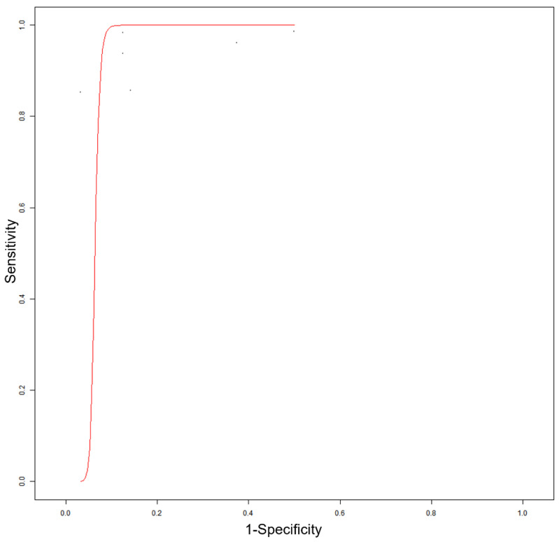
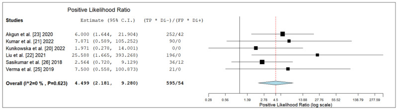
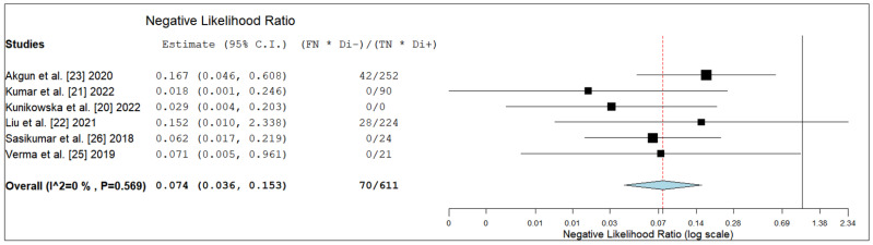
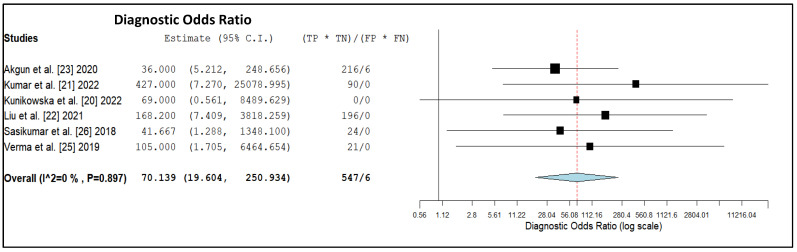
Results: The meta-analysis of six selected studies (157 patients) provided the following results about PET/CT or PET/MRI with PSMA-targeting radiopharmaceuticals in the diagnosis of HGG: sensitivity 98.2% (95% CI: 75.3-99.9%), specificity 91.2% (95% CI: 68.4-98.1%), LR+ 4.5 (95% CI: 2.2-9.3), LR- 0.07 (95% CI: 0.04-0.15), and DOR 70.1 (95% CI: 19.6-250.9). No significant statistical heterogeneity among the included studies was found (I2 = 0%).
Conclusions: the quantitative data provided demonstrate the high diagnostic accuracy of PET/CT or PET/MRI with PSMA-targeting radiopharmaceuticals for HGG detection. However, more studies are needed to confirm the promising role of PSMA-targeted PET in this clinical setting.
Keywords: PET; PSMA; brain tumors; glioblastoma; glioma; meta-analysis; neuro-oncology; nuclear medicine; positron emission tomography.
Conflict of interest statement
Outside the submitted work: JK reports an unrestricted grant from Janssen, consulting fees from Telix and Novartis. The other coauthors declare no conflicts of interest.
Figures
References
-
- Weller M., van den Bent M., Preusser M., Le Rhun E., Tonn J.C., Minniti G., Bendszus M., Balana C., Chinot O., Dirven L., et al. EANO guidelines on the diagnosis and treatment of diffuse gliomas of adulthood. Nat. Rev. Clin. Oncol. 2021;18:170–186. doi: 10.1038/s41571-020-00447-z. - DOI - PMC - PubMed
-
- Law I., Albert N.L., Arbizu J., Boellaard R., Drzezga A., Galldiks N., la Fougère C., Langen K.J., Lopci E., Lowe V., et al. Joint EANM/EANO/RANO practice guidelines/SNMMI procedure standards for imaging of gliomas using PET with radiolabelled amino acids and [18F]FDG: Version 1.0. Eur. J. Nucl. Med. Mol. Imaging. 2019;46:540–557. doi: 10.1007/s00259-018-4207-9. - DOI - PMC - PubMed
Publication types
LinkOut - more resources
Full Text Sources
Miscellaneous
