Review
. 2022 Jul 12;12(7):1693.
doi: 10.3390/diagnostics12071693.

Extra-Gynecological Pelvic Pathology: A Challenge in the Differential Diagnosis of the Female Pelvis

Affiliations
Review

Extra-Gynecological Pelvic Pathology: A Challenge in the Differential Diagnosis of the Female Pelvis

Betlem Graupera et al. Diagnostics (Basel). .

Abstract

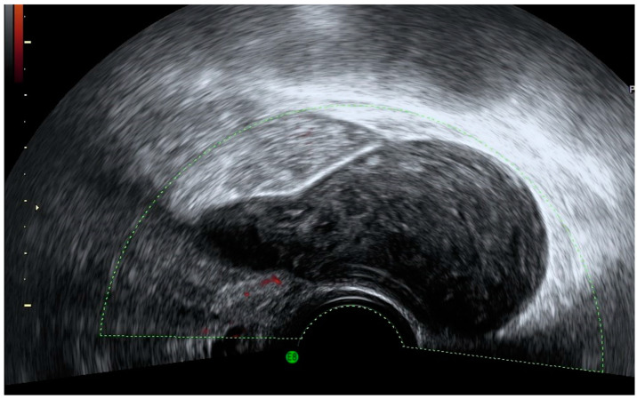
Ultrasound technology with or without color Doppler allows a real-time evaluation of the entire female pelvis including gynecologic and non-gynecological organs, as well as their pathology. As ultrasound is an accurate tool for gynecological diagnosis and is less invasive and less expensive than other techniques, it should be the first imaging modality used in the evaluation of the female pelvis. We present a miscellany of non-gynecological pelvic images observed during the realization of gynecological ultrasound. Transvaginal and transabdominal ultrasound is the first choice among diagnostic techniques for the study of the female pelvis, providing information about gynecological and extra-gynecological organs, allowing for an orientation toward the pathology of a specific organ or system as well as for additional tests to be performed that are necessary for definitive diagnosis.

Keywords: color Doppler; extra-gynecological disease; female pelvis; ultrasound.


Conflict of interest statement

The authors declare no conflict of interest.

Figures

Figure 1
Figure 1
Grayscale transvaginal ultrasound of an inflamed appendix. Longitudinal transvaginal sonographic image demonstrates a blind-ending tubular structure in the right adnexal region pointed with yellow arrow and a small periappendiceal fluid collection (white arrow) (A). Transverse sonographic view showing the right ovary (white arrow) and the characteristic submucosal ring of an inflamed appendix (yellow arrow) (B).
Figure 2
Figure 2
Power Doppler demonstrates hyperemia in the wall of the appendix (black arrow).
Figure 3
Figure 3
Longitudinal transvaginal ultrasound with color Doppler demonstrates the presence of intraluminal gas in the appendix (white arrow) and hyperemia (black arrow).
Figure 4
Figure 4
Grayscale (A) and color Doppler (B) of appendicular plastron showing a vascularized heterogeneous mass in the right iliac fossa.
Figure 5
Figure 5
Power Doppler transvaginal ultrasound shows an appendiceal mucocele as a well-defined, thin-walled cystic mass with pear-shaped morphology with echogenic content and no vascularization.
Figure 6
Figure 6
Sonographic features of an appendiceal mucocele. Ultrasound demonstrates the presence of characteristic concentric echogenic layers (arrows) within a cystic mass or “onion skin” pattern.
Figure 7
Figure 7
A three-dimensional rendering of a mucocele of 96 × 46 × 20 mm in an 80-year-old woman (A) and the same mucocele at surgery (B).
Figure 8
Figure 8
Transvaginal grayscale ultrasound in a patient with acute diverticulitis shows a hyperechoic image arising from the colonic wall with a hypoechoic rim representing wall thickening of the inflamed diverticulum (yellow arrow) and minimal wall thickening of the colon (black arrows).
Figure 9
Figure 9
Transvaginal grayscale (A) and color Doppler image (B) of a patient with colonic carcinoma. Note the focal thickening of the colonic wall (double arrow) and the abundant vascularization in color Doppler study.
Figure 10
Figure 10
Transvaginal (A) and transabdominal (B) ultrasound of a patient with a GIST shows a solid pelvic tumor of mixed echogenicity with cystic areas (yellow arrows) and abundant vascularization and without acoustic shadowing.
Figure 11
Figure 11
Power Doppler transvaginal ultrasound (A) and color Doppler transabdominal ultrasound (B) demonstrate abundant vascularization inside the GIST.
Figure 12
Figure 12
Ultrasound imaging demonstrates a megaureter with tapered distal segment (white arrows) ending at the bladder (yellow arrow) (A). Color Doppler shows no vascularization of megaureter (B).
Figure 13
Figure 13
Ultrasonographic image of ureterocele showing an anechoic cyst (white arrow) within the posterior aspect of the urinary bladder (yellow arrow).
Figure 14
Figure 14
Transvaginal ultrasound shows a ureterocele (yellow arrow). Color Doppler demonstrates the flow of urine (white arrow) toward the urinary bladder (UB).
Figure 15
Figure 15
Transvaginal ultrasound shows the urinary bladder with an irregular solid, and polypoid intravesical lesion between the calipers (A). Power Doppler reveals high vascularization (B). Histology confirmed a bladder carcinoma.
Figure 16
Figure 16
Transvaginal ultrasound shows a transversal view of the uterus (yellow arrow) and a solid mass in the right iliac fossa pointed with white arrows, presenting the ultrasonographic renal characteristics corresponding to an ectopic kidney.
Figure 17
Figure 17
Color Doppler transvaginal ultrasound shows the vascularization of an ectopic kidney (A). Transabdominal ultrasound in the same patient demonstrates the presence of the liver (yellow arrow) and the absence of the orthotopic kidney (B).
Figure 18
Figure 18
Color Doppler transvaginal ultrasound (A,B) demonstrates dilated and tortuous arcuate uterine vessels communicating with varicose pelvic veins. Uterus is indicated with yellow arrow (A).
Figure 19
Figure 19
Grayscale (A) and power Doppler (B) transvaginal ultrasound show a uterine vein with the presence of a thrombus (yellow arrow) inside. Note the absence of vascularization in the image corresponding to the thrombus.
Figure 20
Figure 20
Transvaginal ultrasonography shows a septate cystic nodule independent from ovary corresponding to ovarian vein thrombosis. Power Doppler demonstrates peripheral and central vascularization.
Figure 21
Figure 21
Grayscale transvaginal ultrasound shows a pelvic rounded cystic mass (A). Color Doppler evidence vascularization demonstrates the vascular nature of the lesion corresponding to a pelvic aneurysm (B).
Figure 22
Figure 22
Transvaginal ultrasound shows an irregular nodule with diffuse echoes consisting of lymphoma (A). Color Doppler demonstrates central vascularization (B).
Figure 23
Figure 23
Soft-tissue ultrasound demonstrates an irregular hypoechoic nodule (A) in which power Doppler demonstrates abundant penetrating vascularization (B). Both Figure 22 and Figure 23 depict lymphoma.
Figure 24
Figure 24
(A,B) Cystic lesion related to iliac vessels in a patient with previous lymphadenectomy consistent with lymphocele.
Figure 25
Figure 25
Transvaginal ultrasound shows a pelvic multilocular solid lesion independent from both ovaries in a patient with previous cystic lymphangioma.
Figure 26
Figure 26
Transvaginal ultrasound shows Tarlov cyst as a well-defined cystic lesion in the adnexal region with the presence of some echoes or a network of very fine walls inside it (AD).
Figure 27
Figure 27
Color Doppler transvaginal ultrasound shows in the right adnexal region, a well-defined solid mass with cystic areas and scattered vessels in a patient with histological result of neurofibroma.
Figure 28
Figure 28
Transvaginal ultrasound sagittal view of the pelvis shows the uterus and the cervix and adjacent to the posterior wall, a cystic lesion with diffuse echoes, and hyperechoic images with acoustic shadows consistent with a pilonidal cyst (AC). Power Doppler shows absence of vascularization (C).
Figure 29
Figure 29
Transvaginal ultrasound imaging (A,B) shows a heterogeneous solid mass with acoustic shadows in a patient with recent pelvic surgery. Color Doppler does not demonstrate vascularization (B). This image corresponds to a surgical gossypiboma.

References

    1. Benacerraf B.R., Abuhamad A.Z., Bromley B., Goldstein S.R., Groszmann Y., Shipp T.D., Timor-Tritsch I. Consider ultrasound first for imaging the female pelvis. Am. J. Obstet. Gynecol. 2015;212:450–455. doi: 10.1016/j.ajog.2015.02.015. - DOI - PubMed
    1. Minton K.K., Abuhamad A.Z. 2012 Ultrasound First Forum proceedings. J. Ultrasound Med. 2013;32:555–566. doi: 10.7863/jum.2013.32.4.555. - DOI - PubMed
    1. Loutradis D., Antsaklis A., Creatsas G., Hatzakis A., Kanakas N., Gougoulakis A., Michalas S., Aravantinos D. The validity of gynecological ultrasonography. Gynecol. Obstet. Investig. 1990;29:47–50. doi: 10.1159/000293299. - DOI - PubMed
    1. Beyer D., Schulte B., Kaiser C. Ultrasound diagnosis of the acute abdomen. Bildgebung. 1993;60:241–247. - PubMed
    1. Recker F., Weber E., Strizek B., Gembruch U., Westerway S.C., Dietrich C.F. Point-of-care ultrasound in obstetrics and gynecology. Arch. Gynecol. Obstet. 2021;303:871–876. doi: 10.1007/s00404-021-05972-5. - DOI - PMC - PubMed

LinkOut - more resources