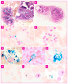
Perls' Stain Guidelines from the French-Speaking Cellular Hematology Group (GFHC)
- PMID: 35885602
- PMCID: PMC9318570
- DOI: 10.3390/diagnostics12071698
Perls' Stain Guidelines from the French-Speaking Cellular Hematology Group (GFHC)
Abstract
In order to standardize cellular hematology practices, the French-speaking Cellular Hematology Group (Groupe Francophone d'Hématologie Cellulaire, GFHC) focused on Perls' stain. A national survey was carried out, leading to the proposal of recommendations on insoluble iron detection and quantification in bone marrow. The criteria presented here met with a "strong professional agreement" and follow the suggestions of the World Health Organization's classification of hematological malignancies.
Keywords: Perls’ stain; Prussian blue stain; bone marrow; cytochemistry; iron staining; iron store; myelodysplastic syndromes; recommendations; sideroblasts.
Conflict of interest statement
The authors declare no conflict of interest.
Figures


References
-
- Swerdlow S.H., Campo E. WHO Classification of Tumors of Haematopoietic and Lymphoid Tissues. IARC revised 4th ed. IARC; Lyon, France: 2017.
-
- Bain B.J., Bates I. Dacie and Lewis Practical Haematology. 12th ed. Elsevier; London, UK: 2017.
-
- Guillosson J.J., Nafziger J. Fiche technique: Recherche des sidéroblastes et sidérocytes dans la moelle osseuse par la réaction de Perls. Feuill. de Biol. Clin. 1987;28:41–42.
Publication types
LinkOut - more resources
Full Text Sources
Miscellaneous

