

Detection of Skin Cancer Based on Skin Lesion Images Using Deep Learning
- PMID: 35885710
- PMCID: PMC9324455
- DOI: 10.3390/healthcare10071183
Detection of Skin Cancer Based on Skin Lesion Images Using Deep Learning
Abstract
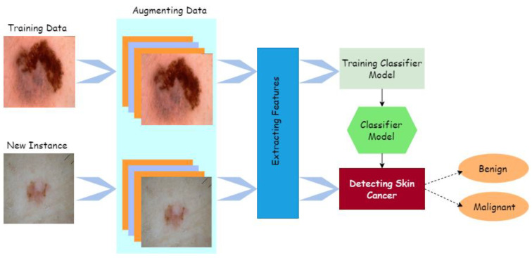
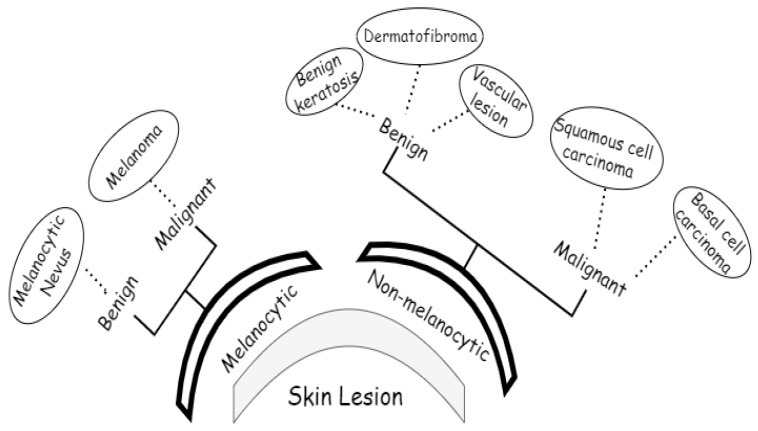
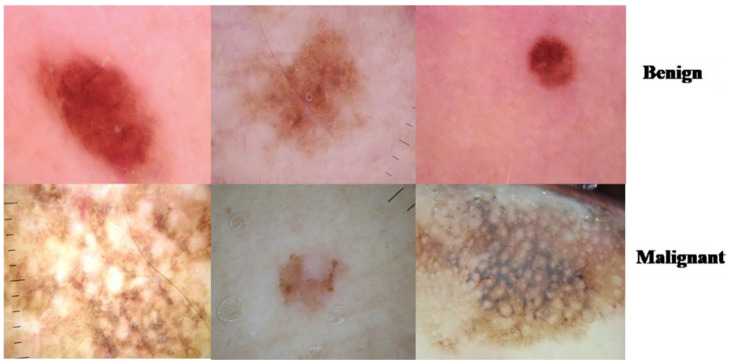
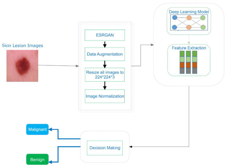
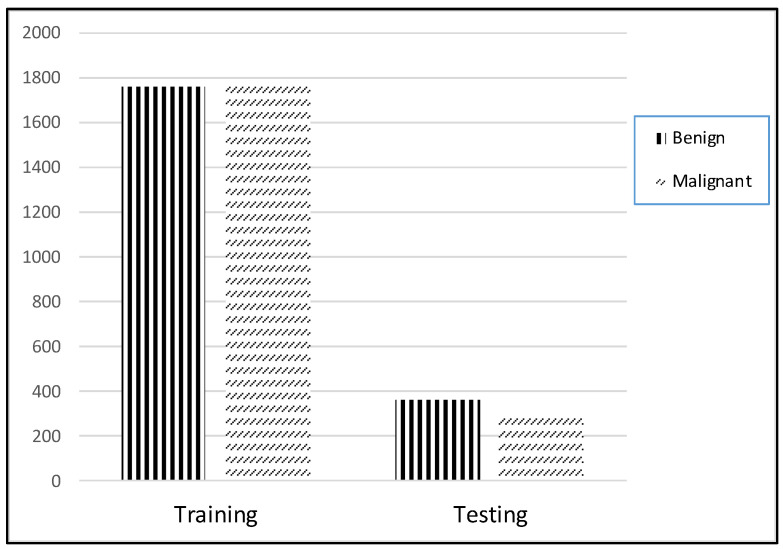
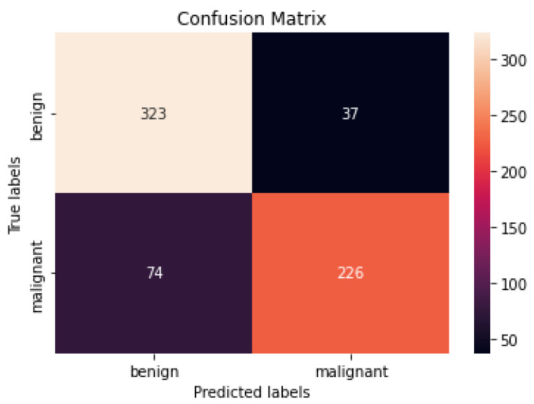
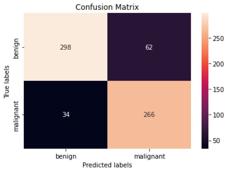
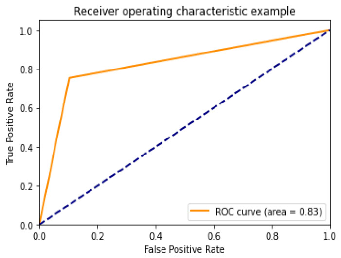
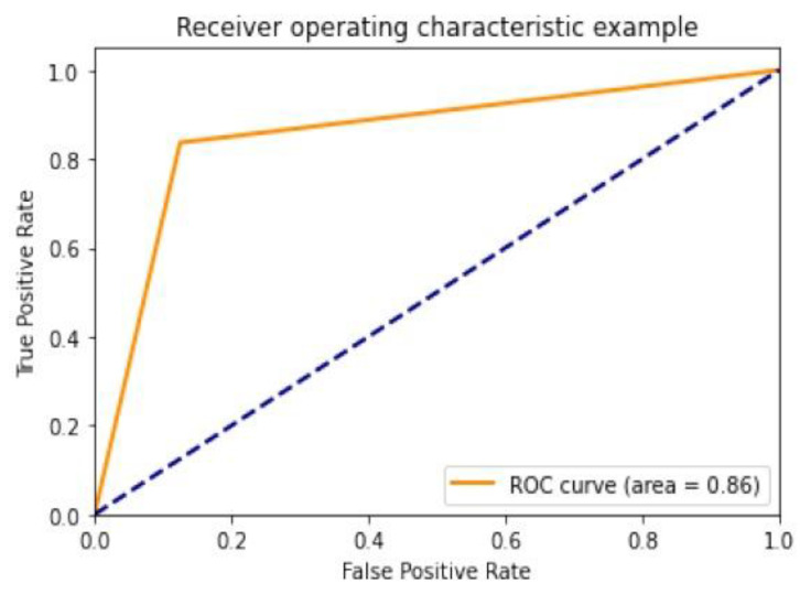
An increasing number of genetic and metabolic anomalies have been determined to lead to cancer, generally fatal. Cancerous cells may spread to any body part, where they can be life-threatening. Skin cancer is one of the most common types of cancer, and its frequency is increasing worldwide. The main subtypes of skin cancer are squamous and basal cell carcinomas, and melanoma, which is clinically aggressive and responsible for most deaths. Therefore, skin cancer screening is necessary. One of the best methods to accurately and swiftly identify skin cancer is using deep learning (DL). In this research, the deep learning method convolution neural network (CNN) was used to detect the two primary types of tumors, malignant and benign, using the ISIC2018 dataset. This dataset comprises 3533 skin lesions, including benign, malignant, nonmelanocytic, and melanocytic tumors. Using ESRGAN, the photos were first retouched and improved. The photos were augmented, normalized, and resized during the preprocessing step. Skin lesion photos could be classified using a CNN method based on an aggregate of results obtained after many repetitions. Then, multiple transfer learning models, such as Resnet50, InceptionV3, and Inception Resnet, were used for fine-tuning. In addition to experimenting with several models (the designed CNN, Resnet50, InceptionV3, and Inception Resnet), this study's innovation and contribution are the use of ESRGAN as a preprocessing step. Our designed model showed results comparable to the pretrained model. Simulations using the ISIC 2018 skin lesion dataset showed that the suggested strategy was successful. An 83.2% accuracy rate was achieved by the CNN, in comparison to the Resnet50 (83.7%), InceptionV3 (85.8%), and Inception Resnet (84%) models.
Keywords: ISIC 2018; computer vision; convolutional neural network; deep learning; machine learning; skin lesion.
Conflict of interest statement
The authors declare no conflict of interest.
Figures

Similar articles
-
Multiple skin lesions diagnostics via integrated deep convolutional networks for segmentation and classification.Comput Methods Programs Biomed. 2020 Jul;190:105351. doi: 10.1016/j.cmpb.2020.105351. Epub 2020 Jan 23. Comput Methods Programs Biomed. 2020. PMID: 32028084
-
Developing an efficient method for melanoma detection using CNN techniques.J Egypt Natl Canc Inst. 2024 Feb 26;36(1):6. doi: 10.1186/s43046-024-00210-w. J Egypt Natl Canc Inst. 2024. PMID: 38407684
-
Enhancing Skin Lesion Detection: A Multistage Multiclass Convolutional Neural Network-Based Framework.Bioengineering (Basel). 2023 Dec 15;10(12):1430. doi: 10.3390/bioengineering10121430. Bioengineering (Basel). 2023. PMID: 38136020 Free PMC article.
-
Deep Learning Approaches Towards Skin Lesion Segmentation and Classification from Dermoscopic Images - A Review.Curr Med Imaging. 2020;16(5):513-533. doi: 10.2174/1573405615666190129120449. Curr Med Imaging. 2020. PMID: 32484086 Review.
-
Multi-features extraction based on deep learning for skin lesion classification.Tissue Cell. 2022 Feb;74:101701. doi: 10.1016/j.tice.2021.101701. Epub 2021 Nov 25. Tissue Cell. 2022. PMID: 34861582 Review.
Cited by
-
A Multi-level ensemble approach for skin lesion classification using Customized Transfer Learning with Triple Attention.PLoS One. 2024 Oct 24;19(10):e0309430. doi: 10.1371/journal.pone.0309430. eCollection 2024. PLoS One. 2024. PMID: 39446759 Free PMC article.
-
Hybrid Deep Learning Framework for Melanoma Diagnosis Using Dermoscopic Medical Images.Diagnostics (Basel). 2024 Oct 8;14(19):2242. doi: 10.3390/diagnostics14192242. Diagnostics (Basel). 2024. PMID: 39410645 Free PMC article.
-
VGG16 Feature Extractor with Extreme Gradient Boost Classifier for Pancreas Cancer Prediction.J Imaging. 2023 Jul 7;9(7):138. doi: 10.3390/jimaging9070138. J Imaging. 2023. PMID: 37504815 Free PMC article.
-
A hybrid parallel convolutional spiking neural network for enhanced skin cancer detection.Sci Rep. 2025 Apr 1;15(1):11137. doi: 10.1038/s41598-025-85627-6. Sci Rep. 2025. PMID: 40169652 Free PMC article.
-
Dual-stage segmentation and classification framework for skin lesion analysis using deep neural network.Digit Health. 2025 Jul 13;11:20552076251351858. doi: 10.1177/20552076251351858. eCollection 2025 Jan-Dec. Digit Health. 2025. PMID: 40666627 Free PMC article.
References
-
- World Health Organization . Global Health Observatory. World Health Organization; Geneva, Switzerland: 2022.
-
- Wolf M., de Boer A., Sharma K., Boor P., Leiner T., Sunder-Plassmann G., Moser E., Caroli A., Jerome N.P. Magnetic resonance imaging T1-and T2-mapping to assess renal structure and function: A systematic review and statement paper. Nephrol. Dial. Transplant. 2018;33((Suppl. S2)):ii41–ii50. doi: 10.1093/ndt/gfy198. - DOI - PMC - PubMed
LinkOut - more resources
Full Text Sources
