. 2022 Jul 7;23(14):7547.
doi: 10.3390/ijms23147547.

Tumor-Localized Administration of α-GalCer to Recruit Invariant Natural Killer T Cells and Enhance Their Antitumor Activity against Solid Tumors

Affiliations

Tumor-Localized Administration of α-GalCer to Recruit Invariant Natural Killer T Cells and Enhance Their Antitumor Activity against Solid Tumors

Yan-Ruide Li et al. Int J Mol Sci. .

Abstract

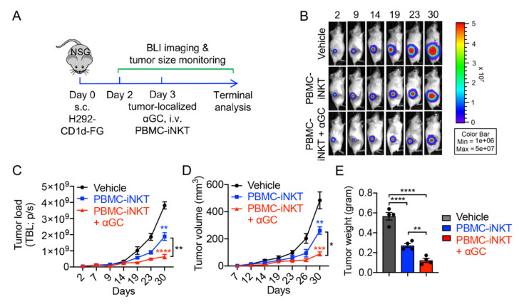
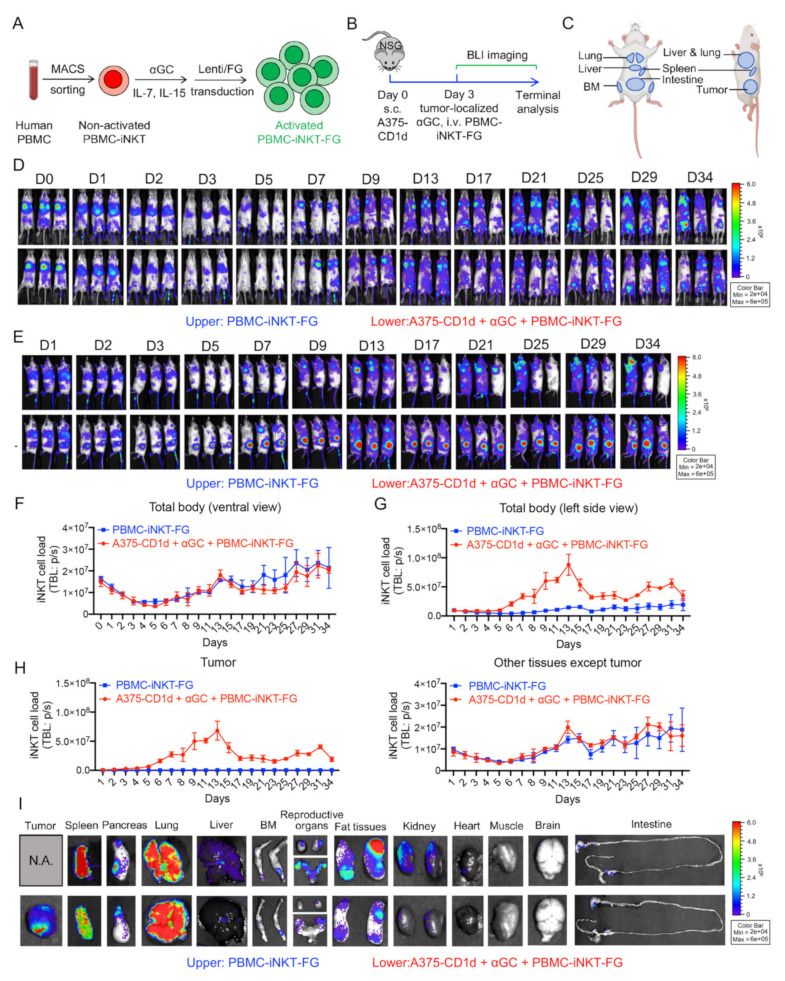
Invariant natural killer T (iNKT) cells have the capacity to mount potent anti-tumor reactivity and have therefore become a focus in the development of cell-based immunotherapy. iNKT cells attack tumor cells using multiple mechanisms with a high efficacy; however, their clinical application has been limited because of their low numbers in cancer patients and difficulties in infiltrating solid tumors. In this study, we aimed to overcome these critical limitations by using α-GalCer, a synthetic glycolipid ligand specifically activating iNKT cells, to recruit iNKT to solid tumors. By adoptively transferring human iNKT cells into tumor-bearing humanized NSG mice and administering a single dose of tumor-localized α-GalCer, we demonstrated the rapid recruitment of human iNKT cells into solid tumors in as little as one day and a significantly enhanced tumor killing ability. Using firefly luciferase-labeled iNKT cells, we monitored the tissue biodistribution and pharmacokinetics/pharmacodynamics (PK/PD) of human iNKT cells in tumor-bearing NSG mice. Collectively, these preclinical studies demonstrate the promise of an αGC-driven iNKT cell-based immunotherapy to target solid tumors with higher efficacy and precision.

Keywords: CD1d; bioluminescence live animal imaging (BLI); cancer immunotherapy; invariant natural killer T (iNKT) cell; lung cancer; melanoma; solid tumor; α-GalCer.


Conflict of interest statement

Lili Yang is a scientific advisor to AlzChem and Amberstone Biosciences, as well as a co-founder, stockholder, and advisory board member of Appia Bio. None of the declared companies contributed to or directed any of the research reported in this article. The remaining authors declare no competing interests.

Figures

Figure 1
Figure 1
Generation of healthy donor peripheral blood mononuclear (PBMC)-derived iNKT (PBMC-iNKT) cells. (A) Experimental design to generate PBMC-iNKT cells in vitro. MACS, magnetic-activated cell sorting; αGC, α-galactosylceramide. (B) PBMC-iNKT cell growth curve post-antigen stimulation. PBMC-iNKT cells were cultured for 13 days in the presence or absence of αGC (denoted as αGC or Vehicle, respectively; n = 3). (C) FACS detection of surface markers on PBMC-iNKT cells. Non-activated PBMC-iNKT cells and non-activated PBMC-Tc cells were included as controls. Representative of three experiments. Data are presented as the mean ± SEM. ** p < 0.01; *** p < 0.001 by Student’s t test.
Figure 2
Figure 2
In vitro tumor cell killing efficacy of PBMC-iNKT cells. (A) Experimental design. Human melanoma A375-CD1d-FG and lung cancer H292-CD1d FG were studied. The two cell lines were generated by engineering parental cell lines to overexpress human CD1d, as well as firefly luciferase and enhanced green fluorescence protein (FG) dual-reporters. (B) Schematics showing the engineered A375-CD1d-FG and H292-CD1d-FG cell lines. (C) FACS detection of CD1d on A375-CD1d-FG and H292-CD1d-FG cells. (D) Tumor killing data at 24 h (n = 4). (E) FACS characterization of CD69, perforin, and granzyme B expression of PBMC-iNKT cells. (F) Quantification of (E) (n = 3). Representative of 3 experiments. Data are presented as the mean ± SEM. ns, not significant, * p < 0.05; *** p < 0.001; **** p < 0.0001 by one-way ANOVA.
Figure 3
Figure 3
In vivo anti-tumor efficacy of PBMC-iNKT cells in an H292-CD1d-FG human lung cancer xenograft NSG mouse model. (A) Experimental design. BLI, live animal bioluminescence imaging; s.c., subcutaneous injection; i.v., intravenous injection. (B) BLI images showing tumor loads in experimental mice over time. (C) Quantification of (B) (n = 4). (D) Tumor size measurements over time (n = 4). (E) Tumor weight measurement on day 33 (n = 4). Representative of 2 experiments. Data are presented as the mean ± SEM. ns, not significant, * p < 0.05; ** p < 0.01; *** p < 0.001; **** p < 0.0001 by one-way ANOVA.
Figure 4
Figure 4
In vivo anti-tumor efficacy of PBMC-iNKT cells in an A375-CD1d-FG human melanoma xenograft NSG mouse model. (A) Experimental design. (B) BLI images showing tumor loads in experimental mice over time. (C) Quantification of (B) (n = 3–4). (D) Tumor size measurements over time (n = 3–4). (E) Tumor weight measurement on day 20 (n = 3–4). Representative of 2 experiments. Data are presented as the mean ± SEM. ns, not significant, * p < 0.05; ** p < 0.01; *** p < 0.001; **** p < 0.0001 by one-way ANOVA.
Figure 5
Figure 5
In vivo pharmacokinetics/pharmacodynamics (PK/PD) study of PBMC-iNKT cells in an A375-CD1d human melanoma xenograft NSG mouse model. (A) Generation of PBMC-iNKT cells overexpressing FG. (B) Experimental design. (C) The templates showing the distribution of tissues in the experimental mice from the ventral and left side views. BM, bone marrow. (D) BLI images showing iNKT cell load in experimental mice over time. The mice were imaged from the ventral view. The upper panel shows the experimental mice injected with PBMC-iNKT-FG cells, and the lower panel shows the experimental mice injected with A375-CD1d tumor cells, αGC, and PBMC-iNKT-FG cells. (E) BLI images showing iNKT cell load in experimental mice over time. The mice were imaged from the left side view. (F) Quantification of (D) (n = 3). (G) Quantification of (E) (n = 3). (H) Quantification of iNKT cell load in tumor and tissues in experimental mice over time. The quantification data of other tissues were obtained by subtracting iNKT cell load in tumor from that in total body of experimental mice. (I) BLI images showing iNKT cell load in multiple tissues from experimental mice on day 35. Representative of 2 experiments.

References

    1. King L.A., Lameris R., de Gruijl T.D., van der Vliet H.J. CD1d-Invariant Natural Killer T Cell-Based Cancer Immunotherapy: α-Galactosylceramide and Beyond. Front. Immunol. 2018;9:1519. doi: 10.3389/fimmu.2018.01519. - DOI - PMC - PubMed
    1. Bendelac A., Savage P.B., Teyton L. The biology of NKT cells. Annu. Rev. Immunol. 2007;25:297–336. doi: 10.1146/annurev.immunol.25.022106.141711. - DOI - PubMed
    1. McEwen-Smith R.M., Salio M., Cerundolo V. The regulatory role of invariant NKT cells in tumor immunity. Cancer Immunol. Res. 2015;3:425–435. doi: 10.1158/2326-6066.CIR-15-0062. - DOI - PMC - PubMed
    1. Li Y.-R., Dunn Z.S., Zhou Y., Lee D., Yang L. Development of Stem Cell-Derived Immune Cells for Off-the-Shelf Cancer Immunotherapies. Cells. 2021;10:3479. doi: 10.3390/cells10123497. - DOI - PMC - PubMed
    1. Li Y.-R., Zhou Y., Kramer A., Yang L. Engineering stem cells for cancer immunotherapy. Trends Cancer. 2021;7:1059–1073. doi: 10.1016/j.trecan.2021.08.004. - DOI - PubMed