. 2022 Jul 10;23(14):7624.
doi: 10.3390/ijms23147624.

Light Triggers the Antiproliferative Activity of Naphthalimide-Conjugated (η6-arene)ruthenium(II) Complexes

Affiliations

Light Triggers the Antiproliferative Activity of Naphthalimide-Conjugated (η6-arene)ruthenium(II) Complexes

Franco Bisceglie et al. Int J Mol Sci. .

Abstract

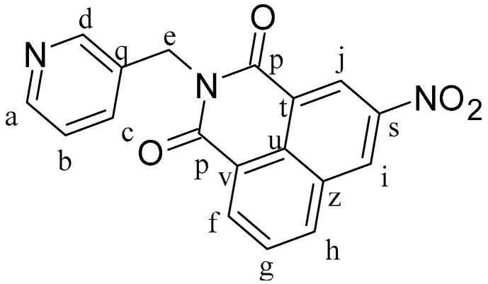
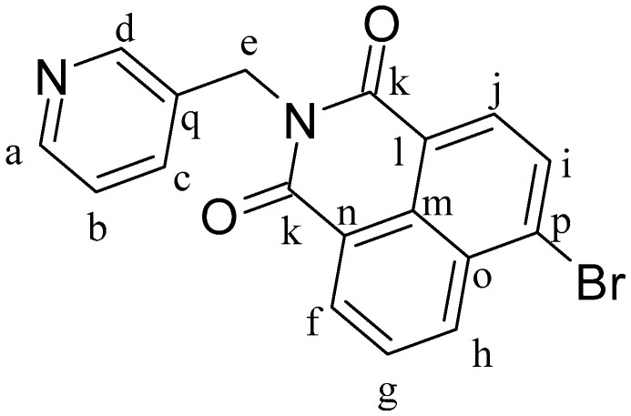
We report the synthesis and characterization of three half-sandwich Ru(II) arene complexes [(η6-arene)Ru(N,N')L][PF6]2 containing arene = p-cymene, N,N' = bipyridine, and L = pyridine meta- with methylenenaphthalimide (C1), methylene(nitro)naphthalimide (C2), or methylene(piperidinyl)naphthalimide (C3). The naphthalimide acts as an antenna for photoactivation. After 3 h of irradiation with blue light, the monodentate pyridyl ligand had almost completely dissociated from complex C3, which contains an electron donor on the naphthalimide ring, whereas only 50% dissociation was observed for C1 and C2. This correlates with the lower wavelength and strong absorption of C3 in this region of the spectrum (λmax = 418 nm) compared with C1 and C2 (λmax = 324 and 323 nm, respectively). All the complexes were relatively non-toxic towards A549 human lung cancer cells in the dark, but only complex C3 exhibited good photocytoxicity towards these cancer cells upon irradiation with blue light (IC50 = 10.55 ± 0.30 μM). Complex C3 has the potential for use in photoactivated chemotherapy (PACT).

Keywords: Ru(II) arene complexes; naphthalimide; photoactivated chemotherapy; photoactivation.


Conflict of interest statement

The authors declare no conflict of interest.

Figures

Figure 1
Figure 1
Structures of the ligands L1, L2, and L3.
Figure 2
Figure 2
Structures of the complexes C1, C2, and C3.
Scheme 1
Scheme 1
Synthetic route used for complexes C1–C3.
Figure 3
Figure 3
UV-vis studies of the stability of solutions of the complexes: (A) C1, (B) C2, and (C) C3, all 20 μM. Comparison between time-zero solution (black line) with the same solution after 24 h of incubation at 37.5 °C in the dark (red line).
Figure 4
Figure 4
Absorption spectra recorded in DMSO at 100 μM of: (A) L1 (black line), L2 (blue line), and L3 (red line); (B) C1 (black line), C2 (blue line), and C3 (red line). Emission spectra in DMSO at 10 μM of: (C) L1 (black line) and C1 (red line), with λex = 370 nm; (D) L2 (black line) and C2 (red line), with λex = 370 nm; and (E) L3 (black line) and C3 (red line), with λex = 418 nm.
Scheme 2
Scheme 2
Photochemical decomposition of the complexes. The color code denotes assignment of peaks for the initial complex (blue) and the products (green and red) in the 1H NMR spectrum (Figure 5, Figure 6 and Figure 7).
Figure 5
Figure 5
1H NMR spectra recorded during the photoactivation of complex C1 with blue light (420 nm) in DMSO-d6.
Figure 6
Figure 6
1H NMR spectra recorded during the photoactivation of complex C2 with blue light (420 nm) in DMSO-d6.
Figure 7
Figure 7
1H NMR spectra recorded during the photoactivation of complex C3 with blue light (420 nm) in DMSO-d6.
Figure 8
Figure 8
Structure of ligand N-(3-methylenepyridyl)-1,8-naphthalimide (L1).
Figure 9
Figure 9
Structure of ligand 3-nitro-N-(3-methylenepyridyl)-1,8-naphthalimide (L2).
Figure 10
Figure 10
Structure of ligand 4-Bromo-N-(3-methylenepyridyl)-1,8-naphthalimide (L1Br).
Figure 11
Figure 11
Structure of ligand 4-Piperidinyl-N-(3-methylenepyridyl)-1,8-naphthalimide (L3).
Figure 12
Figure 12
Structure of complex [(η6-p-cym)Ru(L1)Cl2] (IC1).
Figure 13
Figure 13
Structure of complex [(η6-p-cym)Ru(L2)Cl2] (IC2).
Figure 14
Figure 14
Structure of complex [(η6-p-cym)Ru(L4)Cl2] (IC3).
Figure 15
Figure 15
Structure of complex [(η6-p-cym)Ru(N,N’-bpy)(L1)](SO3CF3)2 (C1).
Figure 16
Figure 16
Structure of complex [(η6-p-cym)Ru(N,N′-bpy)(L2)](SO3CF3)2 (C2).
Figure 17
Figure 17
Structure of complex [(η6-p-cym)Ru(N,N′-bpy)(L4)](SO3CF3)2 (C3).

References

    1. Tandon R., Luxami V., Kaur H., Tandon N., Paul K. 1,8-Naphthalimide: A Potent DNA Intercalator and Target for Cancer Therapy. Chem. Rec. 2017;17:956–993. doi: 10.1002/tcr.201600134. - DOI - PubMed
    1. Monti S., Manet I. Supramolecular photochemistry of drugs in biomolecular environments. Chem. Soc. Rev. 2014;43:4051–4067. doi: 10.1039/C3CS60402K. - DOI - PubMed
    1. Liang G.B., Wei J.H., Jiang H., Huang R.Z., Qin J.T., Wang H.L., Wang H.S., Zhang Y. Design, synthesis and antitumor evaluation of new 1,8-naphthalimide derivatives targeting nuclear DNA. Eur. J. Med. Chem. 2021;210:112951. doi: 10.1016/j.ejmech.2020.112951. - DOI - PubMed
    1. Yin H., Xu Y.F., Qian X.H., Li Y.L., Liu H.W. Novel N-oxide of naphthalimides as prodrug leads against hypoxic solid tumor: Synthesis and biological evaluation. Bioorg. Med. Chem. Lett. 2007;17:2166–2170. doi: 10.1016/j.bmcl.2007.02.015. - DOI - PubMed
    1. Bacchi A., Capucci D., Gatti A., Loffi C., Pioli M., Rogolino D., Terenziani F., Pelagatti P. Synthesis, Structure and Spectroscopic Characterization of Halfsandwich Ru-II Complexes containing 1,8-Naphthalimide Ligands. ChemistrySelect. 2017;2:7000–7007. doi: 10.1002/slct.201701070. - DOI

LinkOut - more resources