

MiTF/TFE Translocation Renal Cell Carcinomas: From Clinical Entities to Molecular Insights
- PMID: 35886994
- PMCID: PMC9324307
- DOI: 10.3390/ijms23147649
MiTF/TFE Translocation Renal Cell Carcinomas: From Clinical Entities to Molecular Insights
Abstract
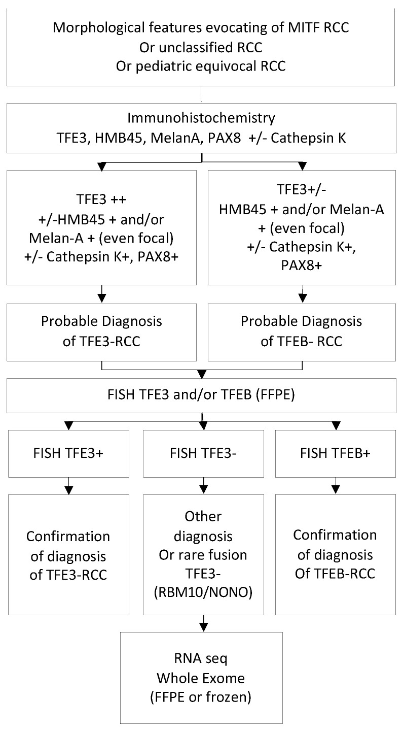
MiTF/TFE translocation renal cell carcinoma (tRCC) is a rare and aggressive subtype of RCC representing the most prevalent RCC in the pediatric population (up to 40%) and making up 4% of all RCCs in adults. It is characterized by translocations involving either TFE3 (TFE3-tRCC), TFEB (TFEB-tRCC) or MITF, all members of the MIT family (microphthalmia-associated transcriptional factor). TFE3-tRCC was first recognized in the World Health Organization (WHO) classification of kidney cancers in 2004. In contrast to TFEB-tRCC, TFE3-tRCC is associated with many partners that can be detected by RNA or exome sequencing. Both diagnoses of TFE3 and TFEB-tRCC are performed on morphological and immunohistochemical features, but, to date, TFE break-apart fluorescent in situ hybridization (FISH) remains the gold standard for diagnosis. The clinical behavior of tRCC is heterogeneous and more aggressive in adults. Management of metastatic tRCC is challenging, especially in the younger population, and data are scarce. Efficacy of the standard of care-targeted therapies and immune checkpoint inhibitors remains low. Recent integrative exome and RNA sequencing analyses have provided a better understanding of the biological heterogeneity, which can contribute to a better therapeutic approach. We describe the clinico-pathological entities, the response to systemic therapy and the molecular features and techniques used to diagnose tRCC.
Keywords: MITF; TFE3; TFEB; translocation renal cell carcinomas.
Conflict of interest statement
The authors declare no conflict of interest.
Figures
References
-
- Durinck S., Stawiski E.W., Pavía-Jiménez A., Modrusan Z., Kapur P., Jaiswal B.S., Zhang N., Toffessi-Tcheuyap V., Nguyen T.T., Pahuja K.B., et al. Spectrum of Diverse Genomic Alterations Define Non–Clear Cell Renal Carcinoma Subtypes. Nat. Genet. 2015;47:13–21. doi: 10.1038/ng.3146. - DOI - PMC - PubMed
-
- Geller J.I., Ehrlich P.F., Cost N.G., Khanna G., Mullen E.A., Gratias E.J., Naranjo A., Dome J.S., Perlman E.J. Characterization of Adolescent and Pediatric Renal Cell Carcinoma: A Report from the Children’s Oncology Group Study AREN03B2. Cancer. 2015;121:2457–2464. doi: 10.1002/cncr.29368. - DOI - PMC - PubMed
Publication types
MeSH terms
Substances
LinkOut - more resources
Full Text Sources
Medical
