Review
. 2022 Jul 12;23(14):7713.
doi: 10.3390/ijms23147713.

PAX Genes in Cardiovascular Development

Affiliations
Review

PAX Genes in Cardiovascular Development

Rebecca E Steele et al. Int J Mol Sci. .

Abstract

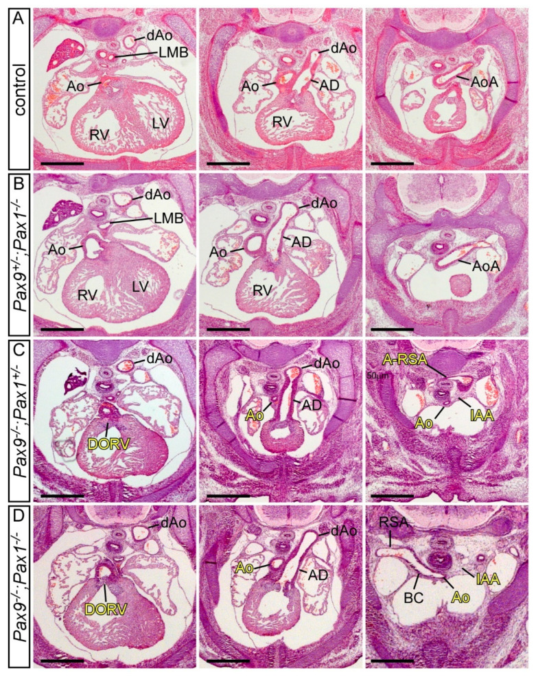
The mammalian heart is a four-chambered organ with systemic and pulmonary circulations to deliver oxygenated blood to the body, and a tightly regulated genetic network exists to shape normal development of the heart and its associated major arteries. A key process during cardiovascular morphogenesis is the septation of the outflow tract which initially forms as a single vessel before separating into the aorta and pulmonary trunk. The outflow tract connects to the aortic arch arteries which are derived from the pharyngeal arch arteries. Congenital heart defects are a major cause of death and morbidity and are frequently associated with a failure to deliver oxygenated blood to the body. The Pax transcription factor family is characterised through their highly conserved paired box and DNA binding domains and are crucial in organogenesis, regulating the development of a wide range of cells, organs and tissues including the cardiovascular system. Studies altering the expression of these genes in murine models, notably Pax3 and Pax9, have found a range of cardiovascular patterning abnormalities such as interruption of the aortic arch and common arterial trunk. This suggests that these Pax genes play a crucial role in the regulatory networks governing cardiovascular development.

Keywords: Pax3; Pax9; cardiovascular development.


Conflict of interest statement

The authors declare no conflict of interest.

Figures

Figure 1
Figure 1
The pharyngeal arches. (A) Mouse embryo at E10.5 with 37 somites (s) with the pharyngeal arches 1–4 labelled. (B) Schematic coronal overview of the tissues and structures of the 3rd, 4th and ultimate (*) pharyngeal arches. Pharyngeal arches are numbered, and pharyngeal pouches (blue arrows) and pharyngeal clefts (pink arrows) indicated. Each pharyngeal arch is filled with neural crest-derived mesenchyme. (C) Coronal section of the embryo in (A) with the section plane shown with a dotted line. (D) Coronal section of an E10.5 embryo immuno-stained with an anti-Pax9 antibody, specifically labelling the pharyngeal endoderm. The 3rd and 4th pharyngeal arch arteries are indicated. Abbreviations: ec, ectoderm; en, endoderm; fl, forelimb bud h, heart; hl, hindlimb bud; paa, pharyngeal arch artery. Scale bar: 500 µm in (A), 100 µm in (C,D).
Figure 2
Figure 2
Development of the aortic arch arteries. Mouse embryos were processed for high resolution episcopic microscopy [9] and the acquired datasets used to make three-dimensional reconstructions with Amira software. (A) At the E10.5 stage the mouse embryo has symmetrical pairs of pharyngeal arch arteries. The first arch artery has remodelled and is no longer visible, the second is interrupted, and the third, fourth and ultimate (*) arch arteries are symmetrical, of equal size and connect the aortic sac (as) with the paired left and right dorsal aorta (lda, rda). The future subclavian arteries, the 7th intersegmental arteries (isa), emanate from the paired dorsal aorta near the point of bifurcation. (B) By E12.5 remodelling of the pharyngeal arch arteries is underway. The outflow tract has separated into the aorta (ao) and pulmonary trunk (pt), the ultimate artery (*) has thinned on the right and expanded on the left. The region of the dorsal aorta between the third and fourth arteries, the carotid duct (cd), is involuting. The right dorsal aorta caudal to the 7th intersegmental artery is regressing. (C) At the fetal stage, E13.5 in the mouse, the arch arteries have completed remodelling to produce the mature aortic arch arteries configuration. (D) Schematic showing the pharyngeal arch arteries (numbered, on the (left)) and the arteries they develop into (labelled, on the (right)). Abbreviations: ao, aorta; as, aortic sac; bc, brachiocephalic artery; cca, common carotid artery; cd, carotid duct; eca, external carotid artery; ica, internal carotid artery; isa, intersegmental artery; lcc, left common carotid; lda, left descending aorta; lsa, left subclavian artery; pa, pulmonary artery; pt, pulmonary trunk; rcc, right common carotid; rda, right descending aorta; rsa, right subclavian artery. Scale bar, 100 µm. Figure adapted from [7].
Figure 3
Figure 3
Cardiovascular developmental defects in Pax9-null mouse embryos. Control (AD) and Pax9-null (Pax9−/−; EH) mouse embryos were collected at different developmental stages and analysed for cardiovascular defects using intracardiac ink injections (A,E), high resolution episcopic microscopy (B,C,F,G) and magnetic resonance imaging (D,H). Control embryos at E10.5-E11.0 have the 3rd, 4th and ultimate (*) arch arteries patent to ink, are symmetrical in appearance and the 1st and 2nd have remodelled at this stage (A,B). In equivalently staged Pax9-null embryos the arch arteries are abnormal, with the 1st and 2nd persisting, the 3rd thin and the 4th absent (E,F). At E11.5 the caudal arch arteries of control embryos start to remodel, with the outflow tract septated and the right ultimate arch artery (*) regressing (C), whereas in Pax9-null embryos outflow tract septation is delayed, the 3rd arch arteries have collapsed and the 1st and/or 2nd persist (G). By E15.5 the pharyngeal arch arteries have remodelled into the adult aortic arch artery configuration (D). Pax9-null embryos show multiple defects (H) including aberrant right subclavian artery (A-RSA), interrupted aortic arch (IAA), abnormal right and left internal and external carotid arteries (i/eRCA, i/eLCA) and double outlet right ventricle (DORV) with interventricular communication (IVC). Somite numbers (s) are indicated. Abbreviations: AD, arterial duct; as, aortic sac; at, aortic trunk; cd, carotid duct; da, dorsal aorta; LCC, left common carotid artery; LSA, left subclavian artery; LV, left ventricle; pa, pulmonary artery; pt, pulmonary trunk; RCC, right common carotid artery; RSA, right subclavian artery; RV, right ventricle. Scale bar: 100 µm in (AC) and (EG), 500 µm in (D,H). Figure adapted from [88].
Figure 4
Figure 4
Developmental defects in Pax9-hypomorphic mouse embryos. Pax9+/Neo control (A,D) and Pax9−/Neo mutant (B,C,E,F) mouse embryos were collected and analysed for cardiovascular defects using magnetic resonance imaging at E15.5 (AC) and intracardiac ink injections at E10.5 (DF). (A) Control embryo with normal cardiovascular anatomy and thymus lobes (white arrows). Pax9−/Neo mutant embryos have normal arch arteries (B) or display fourth pharyngeal arch artery derived defects such as aberrant right subclavian artery and interruption of the aortic arch (C). The thymus lobes are frequently abnormal (yellow arrows; (B,C)). (D) The third (3), fourth (4) and ultimate (*) pharyngeal arch arteries are of equivalent size and patent to ink in control embryos. (E,F) In stage-matched Pax9−/Neo mutant embryos the fourth arch arteries are frequently absent. Somite numbers (s) are indicated. Abbreviations: AD, arterial duct; Ao, aorta; A-RSA, aberrant right subclavian artery; IAA, interrupted aortic arch; RCC, LCC, right/left common carotid artery; RSA, LSA, right/left subclavian artery; RV, LV, right/left ventricle; Tr, trachea. Scale bar: 500 µm in (AC), 100 µm in (DF).
Figure 5
Figure 5
Pax1 and Pax9 do not genetically interact in cardiovascular development. Transverse haematoxylin and eosin-stained sections of E14.5 embryos. (A) Control embryo with normal outflow tract and arch artery morphology with the aorta arising from the left ventricle and the aortic arch crossing over the left main bronchus to join the dorsal aorta. (B) Embryo null for Pax1 heterozygous for Pax9 (Pax9+/−; Pax1−/−) with normal cardiovascular development comparable to the control. (C) Embryo heterozygous for Pax1 and null for Pax9 (Pax9−/−;Pax1+/−) displaying the typical cardiovascular defects seen in Pax9-null embryos. The aorta arises aberrantly from the right ventricle producing a double outlet right ventricle (DORV). The aorta is hypoplastic, with an interruption of the aortic arch (IAA) and an aberrant retro-oesophageal right subclavian artery (A-RSA). (D) Homozygous Pax9;Pax1 null embryo (Pax9−/−;Pax1−/−) also with the typical Pax9-null cardiovascular defects, except with a normal right subclavian artery. Abbreviations: Ao, aorta; AoA, aortic arch; AD, arterial duct; BC, brachiocephalic artery; dAo, dorsal aorta; LMB, left main bronchus; LV, left ventricle; RSA, right subclavian artery; RV, right ventricle. Scale bar: 500 μm.

References

    1. Lough J., Sugi Y. Endoderm and heart development. Dev. Dyn. 2000;217:327–342. doi: 10.1002/(SICI)1097-0177(200004)217:4<327::AID-DVDY1>3.0.CO;2-K. - DOI - PubMed
    1. Stutt N., Song M., Wilson M.D., Scott I.C. Cardiac specification during gastrulation—The Yellow Brick Road leading to Tinman. Semin. Cell Dev. Biol. 2022;127:46–58. doi: 10.1016/j.semcdb.2021.11.011. - DOI - PubMed
    1. Kelly R.G. Molecular Mechanism of Congenital Heart Disease and Pulmonary Hypertension. Springer; Singapore: 2020. Advances in the Second Heart Field; pp. 301–307.
    1. Anderson R.H., Chaudhry B., Mohun T.J., Bamforth S.D., Hoyland D., Phillips H.M., Webb S., Moorman A.F., Brown N.A., Henderson D.J. Normal and abnormal development of the intrapericardial arterial trunks in humans and mice. Cardiovasc. Res. 2012;95:108–115. doi: 10.1093/cvr/cvs147. - DOI - PMC - PubMed
    1. Graham A., Richardson J. Developmental and evolutionary origins of the pharyngeal apparatus. Evodevo. 2012;3:24. doi: 10.1186/2041-9139-3-24. - DOI - PMC - PubMed