

Human Chorionic Villous Differentiation and Placental Development
- PMID: 35887349
- PMCID: PMC9325306
- DOI: 10.3390/ijms23148003
Human Chorionic Villous Differentiation and Placental Development
Abstract
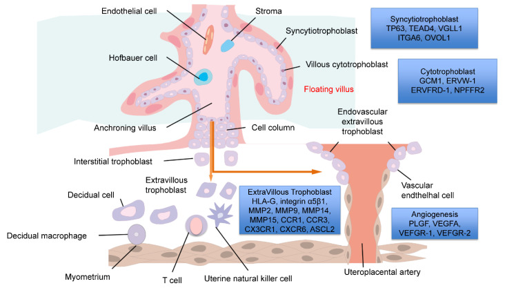
In humans, the placenta provides the only fetomaternal connection and is essential for establishing a pregnancy as well as fetal well-being. Additionally, it allows maternal physiological adaptation and embryonic immunological acceptance, support, and nutrition. The placenta is derived from extra-embryonic tissues that develop rapidly and dynamically in the first weeks of pregnancy. It is primarily composed of trophoblasts that differentiate into villi, stromal cells, macrophages, and fetal endothelial cells (FEC). Placental differentiation may be closely related to perinatal diseases, including fetal growth retardation (FGR) and hypertensive disorders of pregnancy (HDP), and miscarriage. There are limited findings regarding human chorionic villous differentiation and placental development because conducting in vivo studies is extremely difficult. Placental tissue varies widely among species. Thus, experimental animal findings are difficult to apply to humans. Early villous differentiation is difficult to study due to the small tissue size; however, a detailed analysis can potentially elucidate perinatal disease causes or help develop novel therapies. Artificial induction of early villous differentiation using human embryonic stem (ES) cells/induced pluripotent stem (iPS) cells was attempted, producing normally differentiated villi that can be used for interventional/invasive research. Here, we summarized and correlated early villous differentiation findings and discussed clinical diseases.
Keywords: fetal growth retardation; gestational diabetes; hypertensive disorders of pregnancy; iPS cells; placenta; pregnancy.
Conflict of interest statement
The authors declare no conflict of interest.
Figures

References
Publication types
MeSH terms
Grants and funding
LinkOut - more resources
Full Text Sources
Miscellaneous
