Review
. 2022 Jul 21;23(14):8038.
doi: 10.3390/ijms23148038.

Perivascular Mesenchymal Stem/Stromal Cells, an Immune Privileged Niche for Viruses?

Affiliations
Review

Perivascular Mesenchymal Stem/Stromal Cells, an Immune Privileged Niche for Viruses?

Grégorie Lebeau et al. Int J Mol Sci. .

Abstract

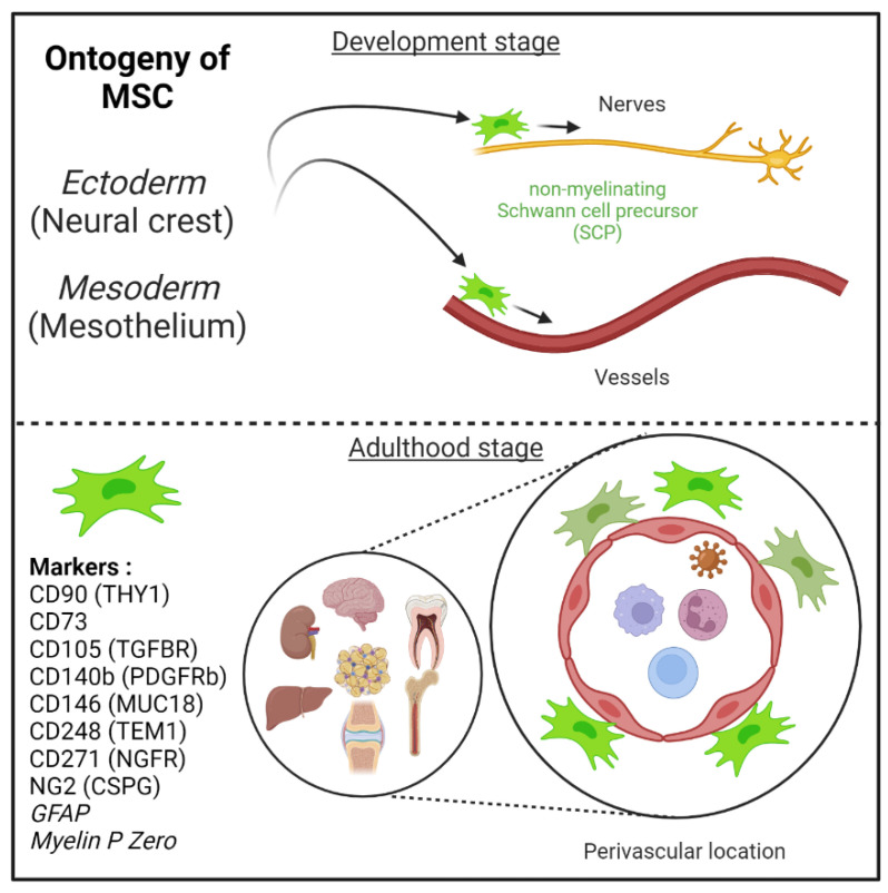
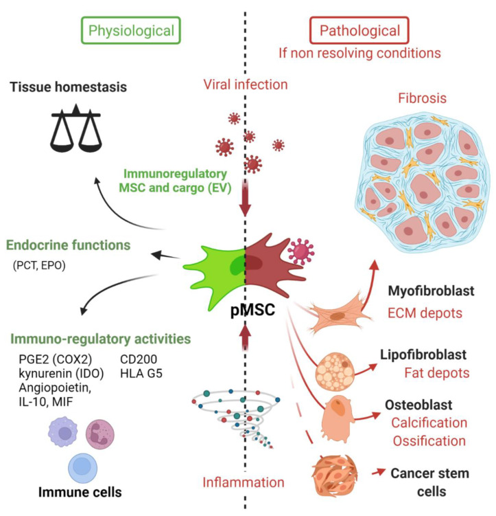
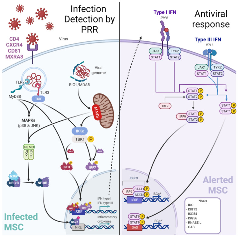
Mesenchymal stem cells (MSCs) play a critical role in response to stress such as infection. They initiate the removal of cell debris, exert major immunoregulatory activities, control pathogens, and lead to a remodeling/scarring phase. Thus, host-derived 'danger' factors released from damaged/infected cells (called alarmins, e.g., HMGB1, ATP, DNA) as well as pathogen-associated molecular patterns (LPS, single strand RNA) can activate MSCs located in the parenchyma and around vessels to upregulate the expression of growth factors and chemoattractant molecules that influence immune cell recruitment and stem cell mobilization. MSC, in an ultimate contribution to tissue repair, may also directly trans- or de-differentiate into specific cellular phenotypes such as osteoblasts, chondrocytes, lipofibroblasts, myofibroblasts, Schwann cells, and they may somehow recapitulate their neural crest embryonic origin. Failure to terminate such repair processes induces pathological scarring, termed fibrosis, or vascular calcification. Interestingly, many viruses and particularly those associated to chronic infection and inflammation may hijack and polarize MSC's immune regulatory activities. Several reports argue that MSC may constitute immune privileged sanctuaries for viruses and contributing to long-lasting effects posing infectious challenges, such as viruses rebounding in immunocompromised patients or following regenerative medicine therapies using MSC. We will herein review the capacity of several viruses not only to infect but also to polarize directly or indirectly the functions of MSC (immunoregulation, differentiation potential, and tissue repair) in clinical settings.

Keywords: COVID-19; chikungunya; chronic inflammation; fibroblast; immune-regulation; immunity; innate immunity; mesenchymal stem cells; neural crest; pericytes; persistence; stromal cells; virus.


Conflict of interest statement

The authors declare no conflict of interest.

Figures

Figure 1
Figure 1
Mesenchymal stem cells (MSC) are derived from either he embryonic ectoderm (neural crest) or the mesoderm. MSCs can migrate along nerves and vessels during development and reside in virtually all post-natal organs and tissues. Along the nerves, MSCs are also known as non-myelinating precursor Schwann cells. Their location around vessels to form perivascular immune privileged niches has been demonstrated by several teams. MSC express several canonical markers which are differentially expressed in all major organs. CD271, GFAP, and MPZ are canonical neuroglial markers.
Figure 2
Figure 2
Deciphering the role of Mesenchymal stem cells in the context of viral infection. In physiological conditions, MSC have important immune functions to control viral infection as glatekeepers around vessels (perivascular MSC) and capable of mounting an innate immune antiviral response. MSC will also promote release of cytokines and chemokines to recruit immune cells to clear pathogens. Equally important is the expression of many immune regulatory factors to terminate the adaptive immune response to limit further cell injuries and promote tissue repair. Many viruses may infect directly MSC in tissues, thus remaining in an immunoprivileged niche favoring virus persistence, spreading and possible virus rebound in immunocompromised patients. Viruses associated to chronic inflammation (non-resolving) may also affect MSC differentiation (e.g., into myofibroblast) leading to excess of extracellular cell matrix production and contributing to organ dysfunction. Importantly, allogenic MSC and derived extracellular vesicles (EV) are nowadays important immunoregulatory cargo injected to patients for the treatment of inflammatory-infectious diseases such as COVID-19. Safety issues are nevertheless highly warranted. PCT (pro-calcitonin); EPO (erythropoietin) hormones.
Figure 3
Figure 3
Infection of MSC by viruses may be controlled by a canonical innate immune response. Viruses may target perivascular MSC naturally expressing receptors (e.g., MXRA8/alphavirus) to grant entry. Among the up-regulated cytosolic pattern recognition receptors (PRRs), Retinoic acid-inducible gene-I (RIG-I)-like receptors (RLR: RIG-I, Melanoma-Differentiation-Associated Gene-5 or MDA5) and Toll-Like Receptor 3 (TLR3) are important to detect viral RNA. After sensing, MSCs engage different cell signaling pathways according to the stimulated PRR. A TLR3-dependent sensing activates mitogen-activated protein kinase pathways (through p38 MAPK and p46 JNK). RLR-dependent sensing stimulates IFN signaling pathway through TBK1/IKK-ε and subsequent interferon regulatory factor (IRF) 7 phosphorylation. These signaling pathways both trigger the production of pro-inflammatory cytokines and peptides with antiviral activities. Hence, in viral context MSCs express increased levels of IL-1β, IL-6, IL-8, IL-11, IL-12p35, IL-23p19, IL-27p28, TNF-α and CCL5/RANTES to recruit and activate adaptive immune cells (T/B lymphocytes). Furthermore, MSCs produce IFN-β and IFN-λ1). Classically, type I IFNs (such as IFN-β) induce the expression of Interferon Stimulated Genes (ISGs, e.g., RNASEL) by the interaction with the IFN receptor (IFNAR).

References

    1. Caplan A. Mesenchymal Stem-Cells. J. Orthop. Res. 1991;9:641–650. doi: 10.1002/jor.1100090504. - DOI - PubMed
    1. Caplan A.I., Dennis J.E. Mesenchymal Stem Cells as Trophic Mediators. J. Cell. Biochem. 2006;98:1076–1084. doi: 10.1002/jcb.20886. - DOI - PubMed
    1. Gomez-Salazar M., Gonzalez-Galofre Z.N., Casamitjana J., Crisan M., James A.W., Peault B. Five Decades Later, Are Mesenchymal Stem Cells Still Relevant? Front. Bioeng. Biotechnol. 2020;8:148. doi: 10.3389/fbioe.2020.00148. - DOI - PMC - PubMed
    1. Da Silva Meirelles L., Caplan A.I., Nardi N.B. In Search of the In Vivo Identity of Mesenchymal Stem Cells. Stem Cells. 2008;26:2287–2299. doi: 10.1634/stemcells.2007-1122. - DOI - PubMed
    1. Crisan M., Yap S., Casteilla L., Chen C.-W., Corselli M., Park T.S., Andriolo G., Sun B., Zheng B., Zhang L., et al. A Perivascular Origin for Mesenchymal Stem Cells in Multiple Human Organs. Cell Stem Cell. 2008;3:301–313. doi: 10.1016/j.stem.2008.07.003. - DOI - PubMed

LinkOut - more resources