Deep Learning to Detect Triangular Fibrocartilage Complex Injury in Wrist MRI: Retrospective Study with Internal and External Validation
- PMID: 35887524
- PMCID: PMC9322609
- DOI: 10.3390/jpm12071029
Deep Learning to Detect Triangular Fibrocartilage Complex Injury in Wrist MRI: Retrospective Study with Internal and External Validation
Abstract
Objective: To use deep learning to predict the probability of triangular fibrocartilage complex (TFCC) injury in patients' MRI scans.
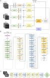
Methods: We retrospectively studied medical records over 11 years and 2 months (1 January 2009-29 February 2019), collecting 332 contrast-enhanced hand MRI scans showing TFCC injury (143 scans) or not (189 scans) from a general hospital. We employed two convolutional neural networks with the MRNet (Algorithm 1) and ResNet50 (Algorithm 2) framework for deep learning. Explainable artificial intelligence was used for heatmap analysis. We tested deep learning using an external dataset containing the MRI scans of 12 patients with TFCC injuries and 38 healthy subjects.
Results: In the internal dataset, Algorithm 1 had an AUC of 0.809 (95% confidence interval-CI: 0.670-0.947) for TFCC injury detection as well as an accuracy, sensitivity, and specificity of 75.6% (95% CI: 0.613-0.858), 66.7% (95% CI: 0.438-0.837), and 81.5% (95% CI: 0.633-0.918), respectively, and an F1 score of 0.686. Algorithm 2 had an AUC of 0.871 (95% CI: 0.747-0.995) for TFCC injury detection and an accuracy, sensitivity, and specificity of 90.7% (95% CI: 0.787-0.962), 88.2% (95% CI: 0.664-0.966), and 92.3% (95% CI: 0.763-0.978), respectively, and an F1 score of 0.882. The accuracy, sensitivity, and specificity for radiologist 1 were 88.9, 94.4 and 85.2%, respectively, and for radiologist 2, they were 71.1, 100 and 51.9%, respectively.
Conclusions: A modified MRNet framework enables the detection of TFCC injury and guides accurate diagnosis.
Keywords: data analysis; deep learning; magnetic resonance imaging; retrospective study; triangular fibrocartilage complex.
Conflict of interest statement
The authors declare no conflict of interest.
Figures


Similar articles
-
Ultrasound With Artificial Intelligence Models Predicted Palmer 1B Triangular Fibrocartilage Complex Injuries.Arthroscopy. 2022 Aug;38(8):2417-2424. doi: 10.1016/j.arthro.2022.03.037. Epub 2022 Apr 18. Arthroscopy. 2022. PMID: 35447195
-
Deep-learning-assisted diagnosis for knee magnetic resonance imaging: Development and retrospective validation of MRNet.PLoS Med. 2018 Nov 27;15(11):e1002699. doi: 10.1371/journal.pmed.1002699. eCollection 2018 Nov. PLoS Med. 2018. PMID: 30481176 Free PMC article.
-
Traumatic injury of the triangular fibrocartilage complex (TFCC)-a refinement to the Palmer classification by using high-resolution 3-T MRI.Skeletal Radiol. 2020 Oct;49(10):1567-1579. doi: 10.1007/s00256-020-03438-4. Epub 2020 May 5. Skeletal Radiol. 2020. PMID: 32372253
-
MR Imaging of the Triangular Fibrocartilage Complex.Magn Reson Imaging Clin N Am. 2015 Aug;23(3):393-403. doi: 10.1016/j.mric.2015.04.001. Magn Reson Imaging Clin N Am. 2015. PMID: 26216770 Review.
-
The Traumatized TFCC: An Illustrated Review of the Anatomy and Injury Patterns of the Triangular Fibrocartilage Complex.Curr Probl Diagn Radiol. 2016 Jan-Feb;45(1):39-50. doi: 10.1067/j.cpradiol.2015.05.004. Epub 2015 May 28. Curr Probl Diagn Radiol. 2016. PMID: 26117527 Review.
Cited by
-
Artificial intelligence in musculoskeletal imaging: realistic clinical applications in the next decade.Skeletal Radiol. 2024 Sep;53(9):1849-1868. doi: 10.1007/s00256-024-04684-6. Epub 2024 Jun 20. Skeletal Radiol. 2024. PMID: 38902420 Review.
-
Artificial intelligence powered advancements in upper extremity joint MRI: A review.Heliyon. 2024 Mar 25;10(7):e28731. doi: 10.1016/j.heliyon.2024.e28731. eCollection 2024 Apr 15. Heliyon. 2024. PMID: 38596104 Free PMC article. Review.
References
-
- Brahim A., Jennane R., Riad R., Janvier T., Khedher L., Toumi H., Lespessailles E. A decision support tool for early detection of knee OsteoArthritis using X-ray imaging and machine learning: Data from the OsteoArthritis Initiative. Comput. Med. Imaging Graph. 2019;73:11–18. doi: 10.1016/j.compmedimag.2019.01.007. - DOI - PubMed
-
- Fong R.C., Vedaldi A. Interpretable explanations of black boxes by meaningful perturbation; Proceedings of the 2017 IEEE International Conference on Computer Vision (ICCV); Venice, Italy. 22–29 October 2017; pp. 3429–3437.
LinkOut - more resources
Full Text Sources

