Review
. 2022 Jul 9;11(14):3988.
doi: 10.3390/jcm11143988.

Proximal Median Nerve Compression in the Differential Diagnosis of Carpal Tunnel Syndrome

Affiliations
Review

Proximal Median Nerve Compression in the Differential Diagnosis of Carpal Tunnel Syndrome

Pekka Löppönen et al. J Clin Med. .

Abstract

Carpal tunnel syndrome (CTS) is the most common median nerve compression neuropathy. Its symptoms and clinical presentation are well known. However, symptoms at median nerve distribution can also be caused by a proximal problem. Pronator syndrome (PS) and anterior interosseous nerve syndrome (AINS) with their typical characteristics have been thought to explain proximal median nerve problems. Still, the literature on proximal median nerve compressions (PMNCs) is conflicting, making this classic split too simple. This review clarifies that PMNCs should be understood as a spectrum of mild to severe nerve lesions along a branching median nerve, thus causing variable symptoms. Clear objective findings are not always present, and therefore, diagnosis should be based on a more thorough understanding of anatomy and clinical testing. Treatment should be planned according to each patient's individual situation. To emphasize the complexity of causes and symptoms, PMNC should be named proximal median nerve syndrome.

Keywords: carpal tunnel syndrome; median nerve entrapment; median neuropathy; neuralgic amyotrophy; pronator syndrome.


Conflict of interest statement

The authors declare no conflict of interest.

Figures
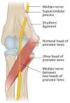
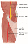
Figure 1
Figure 1
Course of the median nerve at the elbow.
Figure 2
Figure 2
Lacertus fibrosus covers the pronator-flexor mass and the median nerve.
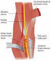
Figure 3
Figure 3
Leading tendinous edge of the flexor digitorum superficialis arch can cause compression of the median nerve and anterior interosseous nerve.
Figure 4
Figure 4
Manual compression of the median nerve at lacertus fibrosus (A) or at pronator teres and FDS arch (B) produces local pain and even distal paresthesia, indicating a nerve compression at that level. Compression test must also be performed at the Struthers’ ligament proximal to the elbow joint level.
Figure 5
Figure 5
FPL and FDP2 weakness in the right hand and inability to make the OK sign indicates nerve injury proximal to the muscles mentioned.
Figure 6
Figure 6
Resisted wrist (A), FDP2 (B,C), and FPL (D) flexion reveals minor weaknesses that the patient might not have yet registered. Weakness in the muscle indicates compression of the median nerve proximal to the site of muscle innervation. All tests must be compared with the unaffected side.
Figure 7
Figure 7
Resisted FDS3 flexion causes pain proximal in the forearm, indicating a nerve compression at the FDS arch.
Figure 8
Figure 8
Resisted elbow flexion tightens the lacertus fibrosus and compresses the median nerve, causing local pain and sometimes distal paresthesia (A). Resisted forearm pronation in full supination tightens the pronator teres and compresses the median nerve (B). Local pain and a loss of pronation power can be observed compared with the unaffected side.
Figure 9
Figure 9
Scratch collapse test. With the patient’s elbows flexed at 90 degrees and the arms held at the sides, the patient externally rotates the arms while the examiner resists the movement (A). After releasing the resistance of the rotation, the skin on top of the median nerve is scratched (B). Instead of scratching the skin, the examiner can compress the median nerve at the point of maximal tenderness (C). After the median nerve irritation, the patient is temporarily unable to resist the rotating force and the affected arm collapses, indicating a proximal compression of the nerve (D).

Similar articles

Cited by

References

    1. Asheghan M., Hollisaz M.T., Aghdam A.S., Khatibiaghda A. The Prevalence of Pronator Teres among Patients with Carpal Tunnel Syndrome: Cross-Sectional Study. Int. J. Biomed. Sci. 2016;12:89–94. - PMC - PubMed
    1. Hsiao C.-W., Shih J.-T., Hung S.-T. Concurrent Carpal Tunnel Syndrome and Pronator Syndrome: A Retrospective Study of 21 Cases. Orthop. Traumatol. Surg. Res. 2017;103:101–103. doi: 10.1016/j.otsr.2016.10.009. - DOI - PubMed
    1. Mujadzic M., Papanicolaou G., Young H., Tsai T.-M. Simultaneous Surgical Release of Ipsilateral Pronator Teres and Carpal Tunnel Syndromes. Plast. Reconstr. Surg. 2007;119:2141–2147. doi: 10.1097/01.prs.0000260703.56453.06. - DOI - PubMed
    1. Olehnik W.K., Manske P.R., Szerzinski J. Median Nerve Compression in the Proximal Forearm. J. Hand Surg. Am. 1994;19:121–126. doi: 10.1016/0363-5023(94)90235-6. - DOI - PubMed
    1. Luangjarmekorn P., Tsai T.M., Honsawek S., Kitidumrongsook P. Role of Pronator Release in Revision Carpal Tunnel Surgery. SICOT J. 2016;2:9. doi: 10.1051/sicotj/2016006. - DOI - PMC - PubMed