

Towards Accurate and Precise Image-Guided Radiotherapy: Clinical Applications of the MR-Linac
- PMID: 35887808
- PMCID: PMC9324978
- DOI: 10.3390/jcm11144044
Towards Accurate and Precise Image-Guided Radiotherapy: Clinical Applications of the MR-Linac
Abstract
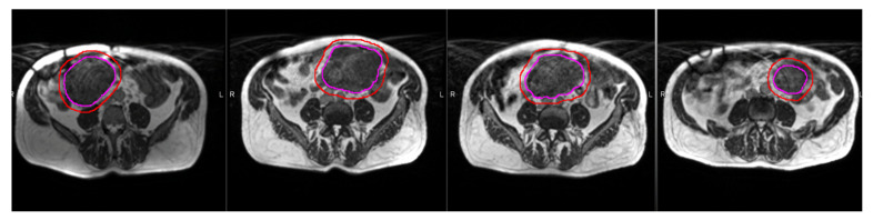
Advances in image-guided radiotherapy have brought about improved oncologic outcomes and reduced toxicity. The next generation of image guidance in the form of magnetic resonance imaging (MRI) will improve visualization of tumors and make radiation treatment adaptation possible. In this review, we discuss the role that MRI plays in radiotherapy, with a focus on the integration of MRI with the linear accelerator. The MR linear accelerator (MR-Linac) will provide real-time imaging, help assess motion management, and provide online adaptive therapy. Potential advantages and the current state of these MR-Linacs are highlighted, with a discussion of six different clinical scenarios, leading into a discussion on the future role of these machines in clinical workflows.
Keywords: IGRT; MRgRT; adaptive therapy; image-guided radiotherapy; linear accelerator; magnetic resonance; radiotherapy.
Conflict of interest statement
The authors declare no conflict of interest.
Figures
References
-
- Henke L., Kashani R., Robinson C., Curcuru A., DeWees T., Bradley J., Green O., Michalski J., Mutic S., Parikh P., et al. Phase I trial of stereotactic MR-guided online adaptive radiation therapy (SMART) for the treatment of oligometastatic or unresectable primary malignancies of the abdomen. Radiother. Oncol. 2018;126:519–526. doi: 10.1016/j.radonc.2017.11.032. - DOI - PubMed
-
- Rosenberg S.A., Henke L.E., Shaverdian N., Mittauer K., Wojcieszynski A.P., Hullett C.R., Kamrava M., Lamb J., Cao M., Green O.L., et al. A Multi-Institutional Experience of MR-Guided Liver Stereotactic Body Radiation Therapy. Adv. Radiat. Oncol. 2019;4:142–149. doi: 10.1016/j.adro.2018.08.005. - DOI - PMC - PubMed
-
- Henke L.E., Olsen J.R., Contreras J.A., Curcuru A., DeWees T.A., Green O.L., Michalski J., Mutic S., Roach M.C., Bradley J.D., et al. Stereotactic MR-Guided Online Adaptive Radiation Therapy (SMART) for Ultracentral Thorax Malignancies: Results of a Phase 1 Trial. Adv. Radiat. Oncol. 2019;4:201–209. doi: 10.1016/j.adro.2018.10.003. - DOI - PMC - PubMed
-
- Bruynzeel A.M.E., Tetar S.U., Oei S.S., Senan S., Haasbeek C.J.A., Spoelstra F.O.B., Piet A.H.M., Meijnen P., Bakker van der Jagt M.A.B., Fraikin T., et al. A Prospective Single-Arm Phase 2 Study of Stereotactic Magnetic Resonance Guided Adaptive Radiation Therapy for Prostate Cancer: Early Toxicity Results. Int. J. Radiat. Oncol. Biol. Phys. 2019;105:1086–1094. doi: 10.1016/j.ijrobp.2019.08.007. - DOI - PubMed
Publication types
LinkOut - more resources
Full Text Sources
