. 2022 Jun 27;12(7):962.
doi: 10.3390/life12070962.

Demyelination Lesions Do Not Correlate with Clinical Manifestations by Bordetella pertussis Toxin Concentrations

Affiliations

Demyelination Lesions Do Not Correlate with Clinical Manifestations by Bordetella pertussis Toxin Concentrations

Maiara Carolina Perussolo et al. Life (Basel). .

Abstract

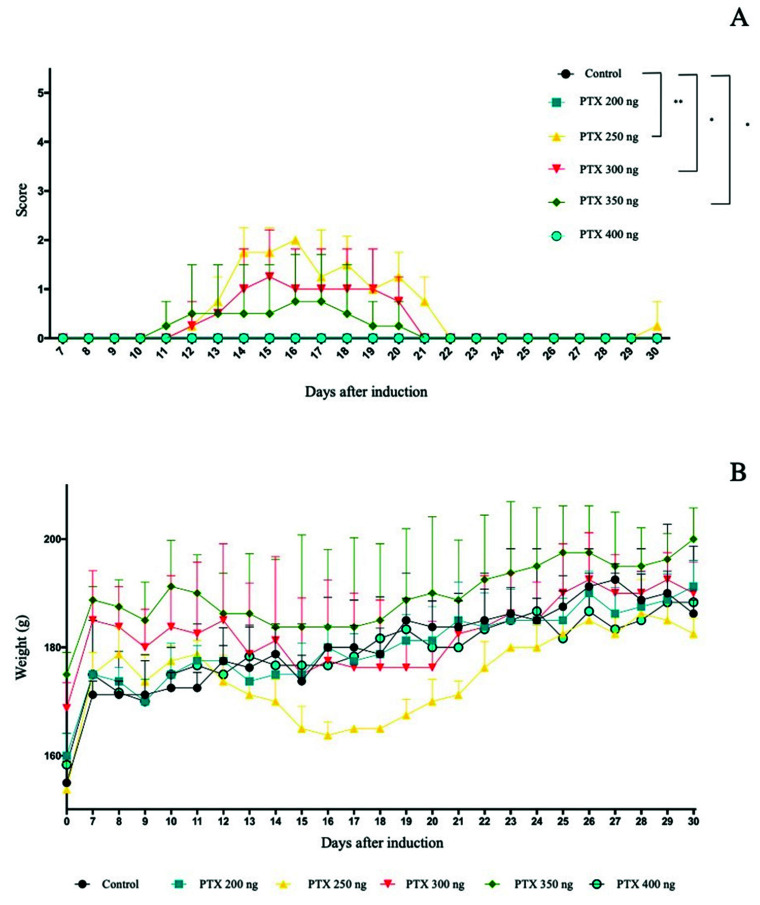
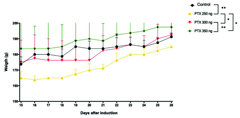
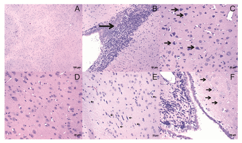
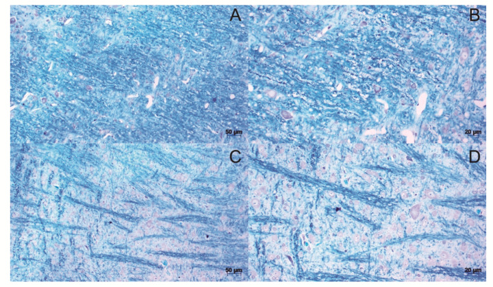
Multiple sclerosis (MS) is an autoimmune disease of the central nervous system, characterized as an inflammatory demyelinating disease. Given the need for improvements in MS treatment, many studies are mainly conducted through preclinical models such as experimental allergic encephalomyelitis (EAE). This study analyzes the relationships between histopathological and clinical score findings at EAE. Twenty-three female Rattus norvegicus Lewis rats from 6 to 8 weeks were induced to EAE. Nineteen rats underwent EAE induction distributed in six groups to establish the evolution of clinical signs, and four animals were in the control group. Bordetella pertussis toxin (PTX) doses were 200, 250, 300, 350 and 400 ng. The clinical scores of the animals were analyzed daily, from seven to 24 days after induction. The brains and spinal cords were collected for histopathological analyses. The results demonstrated that the dose of 250 ng of PTX induced a higher clinical score and reduction in weight. All induced groups demonstrated leukocyte infiltration, activation of microglia and astrocytes, and demyelinated plaques in the brains in histopathology. It was concluded that the dose of 250 ng and 350 ng of PTX were the best choices to trigger the brain and spinal cord demyelination lesions and did not correlate with clinical scores.

Keywords: Bordetella pertussis toxin; animal model; clinical scores; experimental allergic encephalomyelitis; histopathological; multiple sclerosis.


Conflict of interest statement

The abstract of this paper was presented at the Conference Annual Meeting 2021, Coimbra Health School, as an online and oral presentation/conference talk with interim findings. The oral presentation’s abstract was published as “Oral Abstracts” in European Journal of Public Health, Volume 31, Issue Supplement_2, August 2021, ckab120.080: Hyperlink with https://doi.org/10.1093/eurpub/ckab120.080, accessed date 22 June 2022. The authors declare no conflict of interest.

Figures

Figure 1
Figure 1
Evaluation of the score of clinical signs of EAE. Results are expressed as median. Statistical differences when p < 0.05 (*) and p < 0.001 (**). (A); weight variation (g) during the experiment days. Results expressed as mean ± standard error (B).
Figure 2
Figure 2
Analysis of weight (g) of the control, PTX 250, 300, and 350 groups during the peak period and recovery from EAE. Results expressed as mean ± standard error, p < 0.05 (*) and p < 0.001 (**).
Figure 3
Figure 3
Histological sections of brain stained with H&E. (A,D) Tissue from control animals, with a homogeneous distribution of brain cell types. (B) Leukocyte infiltrations (arrow). (C) Neurons on cell death: Observing neurons accompanied by leukocytes (arrows). (E) Activation of microglia (arrows). (F) Astrocyte activation (arrows). The images were obtained from optical microscopy (Carl Zeiss Microscopy, Zeiss, Jena, Germany). Scale bar, 50 µm.
Figure 4
Figure 4
Neuroinflammation score of brain. Results are shown as mean ± standard error and statistically significant differences occur when p < 0.05 (*).
Figure 5
Figure 5
Histological sections of brain stained with Luxol fast blue. Tissue from control animals, with homogeneous distribution of myelin fibers (A,B). demyelinated plaques, present in the brain of animals induced to the model (C,D). The images were obtained from optical microscopy (Carl Zeiss Microscopy, Zeiss, Jena, Germany). Scale bar, 50 µm.
Figure 6
Figure 6
Histological sections of the Spinal cord. The spinal cord of control animals was stained in HE and Luxol fast blue, respectively (A,B). The spinal cord of animals induced to the EAE model, stained in HE and Luxol fast blue, respectively. It is possible to observe the loosening of the myelinated fibers by increasing the axonal space (C,D). The images were obtained from optical microscopy (Carl Zeiss Microscopy, Zeiss, Jena, Germany). Scale bar, 50 µm.

Similar articles

Cited by

References

    1. Lemus H.N., Warrington A.E., Rodriguez M. Multiple Sclerosis. Neurol. Clin. 2018;36:1–11. doi: 10.1016/j.ncl.2017.08.002. - DOI - PMC - PubMed
    1. Al Jumah M.A., Abumaree M.H. The Immunomodulatory and Neuroprotective Effects of Mesenchymal Stem Cells (MSCs) in Experimental Autoimmune Encephalomyelitis (EAE): A Model of Multiple Sclerosis (MS) Int. J. Mol. Sci. 2012;13:9298–9331. doi: 10.3390/ijms13079298. - DOI - PMC - PubMed
    1. Thompson A.J., Baranzini S.E., Geurts J., Hemmer B., Ciccarelli O. Multiple sclerosis. Lancet. 2018;391:1622–1636. doi: 10.1016/S0140-6736(18)30481-1. - DOI - PubMed
    1. Browne P., Chandraratna D., Angood C., Tremlett H., Baker C., Taylor B.V., Thompson A.J. Atlas of Multiple Sclerosis 2013: A growing global problem with widespread inequity. Neurology. 2014;83:1022–1024. doi: 10.1212/WNL.0000000000000768. - DOI - PMC - PubMed
    1. Waubant E., Lucas R., Mowry E., Graves J., Olsson T., Alfredsson L., Langer-Gould A. Environmental and genetic risk factors for MS: An integrated review. Ann. Clin. Transl. Neurol. 2019;6:1905–1922. doi: 10.1002/acn3.50862. - DOI - PMC - PubMed

LinkOut - more resources