Review
. 2022 Jun 30;12(7):982.
doi: 10.3390/life12070982.

The Surgical Treatment of Osteoarthritis

Affiliations
Review

The Surgical Treatment of Osteoarthritis

Peter Brumat et al. Life (Basel). .

Abstract

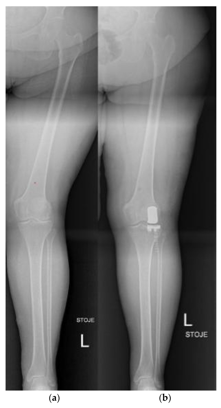
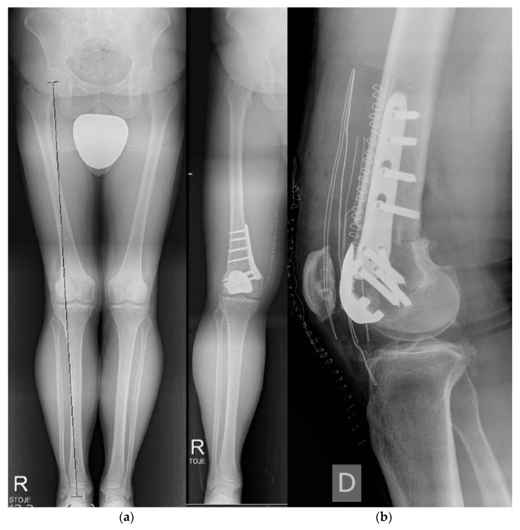
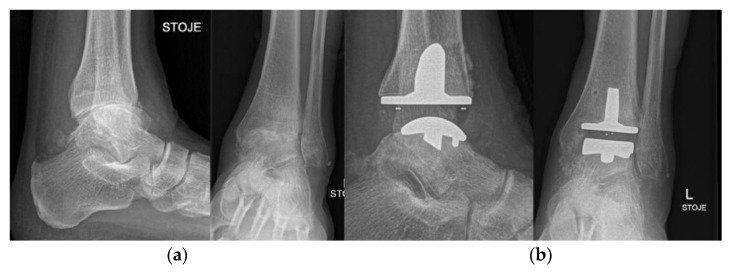
Osteoarthritis is a degenerative condition affecting the whole joint with the underlying bone, representing a major source of pain, disability, and socioeconomic cost worldwide. Age is considered the strongest risk factor, albeit abnormal biomechanics, morphology, congenital abnormality, deformity, malalignment, limb-length discrepancy, lifestyle, and injury may further increase the risk of the development and progression of osteoarthritis as well. Pain and loss of function are the main clinical features that lead to treatment. Although early manifestations of osteoarthritis are amenable to lifestyle modification, adequate pain management, and physical therapy, disease advancement frequently requires surgical treatment. The symptomatic progression of osteoarthritis with radiographical confirmation can be addressed either with arthroscopic interventions, (joint) preservation techniques, or bone fusion procedures, whereas (joint) replacement is preferentially reserved for severe and end-stage disease. The surgical treatment aims at alleviating pain and disability while restoring native biomechanics. Miscellaneous surgical techniques for addressing osteoarthritis exist. Advanced computer-integrated surgical concepts allow for patient personalization and optimization of surgical treatment. The scope of this article is to present an overview of the fundamentals of conventional surgical treatment options for osteoarthritis of the human skeleton, with emphasis on arthroscopy, preservation, arthrodesis, and replacement. Contemporary computer-assisted orthopaedic surgery concepts are further elucidated.

Keywords: CAOS; ankle and foot; computer-assisted orthopaedic surgery; elbow; hip; knee; osteoarthritis; shoulder; spine; wrist and hand.


Conflict of interest statement

The authors declare no conflict of interest. The funders had no role in the design of the study; in the collection, analyses, or interpretation of data; in the writing of the manuscript, or in the decision to publish the results.

Figures

Figure 1
Figure 1
Interspinous spacer. Postoperative X-ray after interspinous spacer implantation (L4–L5 segment).
Figure 2
Figure 2
Interposition (anconeus) arthroplasty. (a,b) preoperative X-rays; (c,d) postoperative X-rays after interposition anconeus arthroplasty.
Figure 3
Figure 3
Interposition (anconeus) arthroplasty, intraoperative images. (a) radial head resection; (b) anconeus muscle preparation; (c) the final position of the interposed anconeus muscle.
Figure 4
Figure 4
Varus femoral osteotomy. (a) preoperative X-ray; (b) postoperative X-ray.
Figure 5
Figure 5
Periacetabular osteotomy (PAO). (a) preoperative X-ray and patient-specific 3D preoperative planning; (b) preoperative 3D CT, planned position of the 3D reoriented acetabular fragment and postoperative X-ray after PAO according to the preoperative patient-specific 3D plan.
Figure 6
Figure 6
Bilateral distal femoral osteotomy (DFO). (a) preoperative X-ray of bilateral limb torsional deformity; (b) postoperative X-ray after bilateral DFO.
Figure 7
Figure 7
Dome-shaped supra-malleolar osteotomy. (a) preoperative X-ray; (b) intraoperative X-ray; (c) postoperative X-ray.
Figure 8
Figure 8
Anterior cervical discectomy and fusion (ACDF). (a) preoperative X-ray; (b) postoperative X-ray of the ACDF (C5–C6 segment).
Figure 9
Figure 9
Shoulder arthrodesis. (a) preoperative X-ray after failed shoulder arthroplasty; (b) postoperative X-ray after shoulder arthrodesis.
Figure 10
Figure 10
Elbow arthrodesis. (a) preoperative X-ray; (b) postoperative X-ray after elbow arthrodesis; (c) intraoperative image.
Figure 11
Figure 11
Secondary conversion of a hip arthrodesis to a total hip arthroplasty. (a) preoperative X-ray after left hip arthrodesis; (b) postoperative X-ray after secondary conversion to a total hip arthroplasty.
Figure 12
Figure 12
Knee arthrodesis. (a) preoperative X-ray in a splint; (b) postoperative X-ray after limb salvage knee arthrodesis with a nail.
Figure 13
Figure 13
Knee arthrodesis. (a) preoperative X-ray of residual knee deformity after severe trauma; (b) postoperative X-ray after limb salvage knee arthrodesis with Ilizarov technique.
Figure 14
Figure 14
Ankle arthrodesis. (a) preoperative X-ray; (b) postoperative X-ray after ankle arthrodesis.
Figure 15
Figure 15
Reverse total shoulder arthroplasty (rTSA). (a) preoperative X-ray; (b) postoperative X-ray after reverse total shoulder arthroplasty.
Figure 16
Figure 16
Total elbow arthroplasty. (a) preoperative X-ray; (b) postoperative X-ray after total elbow arthroplasty.
Figure 17
Figure 17
Bilateral total hip arthroplasty (THA). (a) preoperative X-ray; (b) immediate postoperative X-ray after one-stage bilateral total hip arthroplasty.
Figure 18
Figure 18
Unicompartmental (partial) knee arthroplasty (UKA). (a) preoperative X-ray; (b) postoperative X-ray after unicompartmental knee arthroplasty for isolated knee osteoarthritis of the lateral compartment.
Figure 19
Figure 19
Patellofemoral arthroplasty. (a) preoperative X-ray of isolated patellofemoral osteoarthritis caused by torsional limb deformity; (b) postoperative X-ray after patellofemoral arthroplasty with concomitant distal femoral osteotomy (DFO) for limb deformity correction.
Figure 20
Figure 20
Total ankle arthroplasty (TAA). (a) preoperative X-ray; (b) postoperative X-ray after total ankle arthroplasty.

References

    1. Glyn-Jones S., Palmer A.J.R., Agricola R., Price A.J., Vincent T.L., Weinans H., Carr A.J. Osteoarthritis. Lancet. 2015;386:376–387. doi: 10.1016/S0140-6736(14)60802-3. - DOI - PubMed
    1. Bijlsma J.W., Berenbaum F., Lafeber F.P. Osteoarthritis: An Update with Relevance for Clinical Practice. Lancet. 2011;377:2115–2126. doi: 10.1016/S0140-6736(11)60243-2. - DOI - PubMed
    1. Busija L., Bridgett L., Williams S.R.M., Osborne R.H., Buchbinder R., March L., Fransen M. Osteoarthritis. Best Pract. Res. Clin. Rheumatol. 2010;24:757–768. doi: 10.1016/j.berh.2010.11.001. - DOI - PubMed
    1. Ebell M.H. Osteoarthritis: Rapid Evidence Review. Am. Fam. Physician. 2018;97:523–526. - PubMed
    1. Levesque J.N., Shah A., Ekhtiari S., Yan J.R., Thornley P., Williams D.S. Three-Dimensional Printing in Orthopaedic Surgery: A Scoping Review. EFORT Open Rev. 2020;5:430–441. doi: 10.1302/2058-5241.5.190024. - DOI - PMC - PubMed

LinkOut - more resources