

Non-Destructive Removal of Dental Implant by Using the Cryogenic Method
- PMID: 35888569
- PMCID: PMC9319264
- DOI: 10.3390/medicina58070849
Non-Destructive Removal of Dental Implant by Using the Cryogenic Method
Abstract
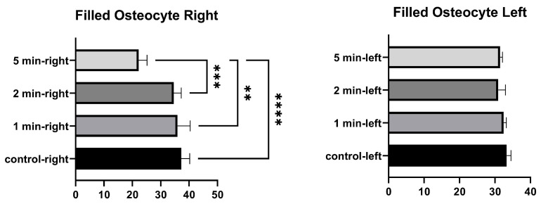
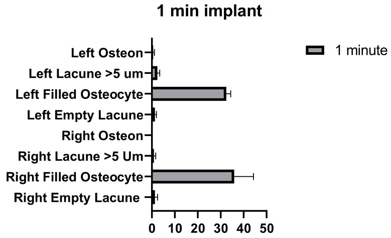
Background and Objectives: The gold standard for a successful prosthetic approach is the osseointegration of an implant. However, this integration can be a problem in cases where the implant needs to be removed. Removing the implant with minimal damage to the surrounding tissues is important. Osteocytes cannot survive below −2 °C, but epithelial cells, fibroblasts, and other surrounding tissue cells can. Remodeling can be triggered by cryotherapy at temperatures that specifically affect osteocyte necrosis. In this study, we aimed to develop a method for reversing the osseointegration mechanism and for protecting the surrounding tissues by bone remodeling induced by CO2 cryotherapy. Materials and Methods: In this study, eight 2.8 mm diameter, one-piece mini implants were used in New Zealand rabbit tibias. Two control and six implants were tested in this study. After 2 months of osseointegration, a reverse torque force method was used to remove all osseointegrated implants at 5, 10, 20, and 30 Ncm. The osseointegration of the implants was proven by periotest measurements. Changes in bone tissue were examined in histological sections stained with toluidine blue after rabbit sacrifice. The number of lacunae with osteocyte, empty lacunae, and lacunae greater than 5 µm and the osteon number in a 10,000 µm2 area were calculated. Cryotherapy was applied to the test implants for 1 min, 2 min, and 5 min. Three implants were subjected to cryotherapy at −40 °C, and the other implants were subjected to cryotherapy at −80 °C. Results: Empty lacunae, filled osteocytes, lacunae >5 µm, and the osteon count around the implant applied at −40 °C were not significantly different from the control implants. The application of −40 °C for 1 min was found to cause minimal damage to the bone cells. The implants, which were applied for 1 min and 2 min, were successfully explanted on the 2nd day with the 5 Ncm reverse torque method. Test implants, which were applied cold for 5 min, were explanted on day 1. Tissue damage was detected in all test groups at −80 °C. Conclusions: The method of removing implants with cryotherapy was found to be successful in −40 °C freeze−thaw cycles applied three times for 1 min. To prove implant removal with cryotherapy, more implant trials should be conducted.
Keywords: carbon dioxide; cryotherapy; implant removal; rabbit; reverse torque; tibia.
Conflict of interest statement
The authors declare no conflict of interest.
Figures


References
-
- Antonelli A., Bennardo F., Brancaccio Y., Barone S., Femiano F., Nucci L., Minervini G., Fortunato L., Attanasio F., Giudice A. Can Bone Compaction Improve Primary Implant Stability? An In Vitro Comparative Study with Osseodensification Technique. Appl. Sci. 2020;10:8623. doi: 10.3390/app10238623. - DOI
-
- Sennerby L., Pagliani L., Petersson A., Verrocchi D., Volpe S., Andersson P. Two different implant designs and impact of related drilling protocols on primary stability in different bone densities: An in vitro comparison study. Int. J. Oral Maxillofac. Implant. 2015;30:564–568. doi: 10.11607/jomi.3903. - DOI - PubMed
MeSH terms
Substances
LinkOut - more resources
Full Text Sources
