

Construction of HGF-Displaying Yeast by Cell Surface Engineering
- PMID: 35889092
- PMCID: PMC9316346
- DOI: 10.3390/microorganisms10071373
Construction of HGF-Displaying Yeast by Cell Surface Engineering
Abstract
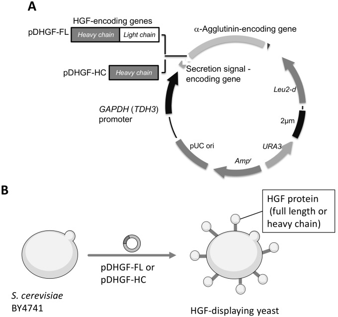
Hepatocyte growth factor (HGF) has been investigated as a regulator for immune reactions caused by transplantation and autoimmune diseases and other biological functions. Previous studies demonstrated that cDNA-encoding HGF administration could inhibit acute graft-versus-host disease (GVHD) after treatment via hematopoietic stem cell transplantation. This study aimed to show the preparation of HGF protein on yeast cell surfaces to develop a tool for the oral administration of HGF to a GVHD mouse model. In this study, full-length HGF and the heavy chain of HGF were genetically fused with α-agglutinin and were successfully displayed on the yeast cell surface. This study suggested that yeast cell surface display engineering could provide a novel administration route for HGF.
Keywords: cell surface; graft versus host disease; hepatocyte growth factor-displaying; oral administration; yeast cells.
Conflict of interest statement
The authors declare no conflict of interest.
Figures


References
-
- Miyazawa K., Shimomura T., Kitamura A., Kondo J., Morimoto Y., Kitamura N. Molecular Cloning and Sequence Analysis of the cDNA for a Human Serine Protease Responsible for Activation of Hepatocyte Growth Factor. Structural Similarity of the Protease Precursor to Blood Coagulation Factor XII. J. Biol. Chem. 1993;268:10024–10028. doi: 10.1016/S0021-9258(18)82167-6. - DOI - PubMed
-
- Chaudhary P., Malhotra S.S., Babu G.S., Sobti R.C., Gupta S.K. HGF Promotes HTR-8/SVneo Cell Migration Through Activation of MAPK/PKA Signaling Leading to Up-Regulation of WNT Ligands and Integrins That Target β-Catenin. Mol. Cell. Biochem. 2019;453:11–32. doi: 10.1007/s11010-018-3428-3. - DOI - PubMed
LinkOut - more resources
Full Text Sources
