. 2022 Jul 7;10(7):1374.
doi: 10.3390/microorganisms10071374.

Comparative Genomic Analysis of the Marine Cyanobacterium Acaryochloris marina MBIC10699 Reveals the Impact of Phycobiliprotein Reacquisition and the Diversity of Acaryochloris Plasmids

Affiliations

Comparative Genomic Analysis of the Marine Cyanobacterium Acaryochloris marina MBIC10699 Reveals the Impact of Phycobiliprotein Reacquisition and the Diversity of Acaryochloris Plasmids

Haruki Yamamoto et al. Microorganisms. .

Abstract

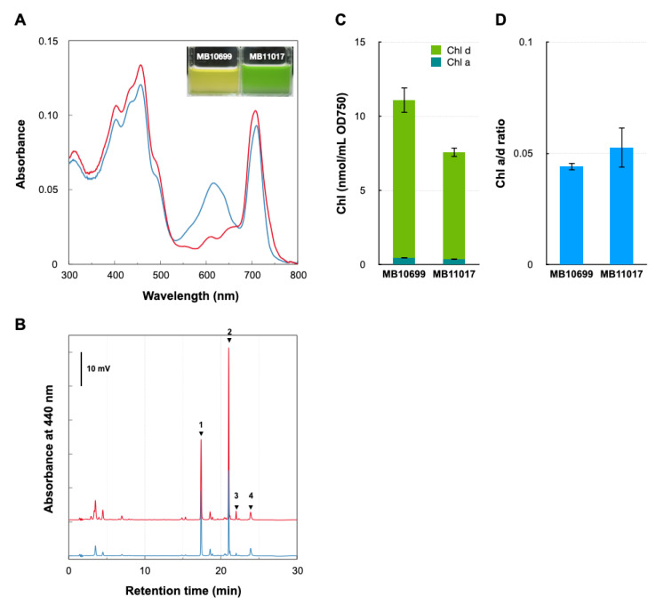
Acaryochloris is a marine cyanobacterium that synthesizes chlorophyll d, a unique chlorophyll that absorbs far-red lights. Acaryochloris is also characterized by the loss of phycobiliprotein (PBP), a photosynthetic antenna specific to cyanobacteria; however, only the type-strain A. marina MBIC11017 retains PBP, suggesting that PBP-related genes were reacquired through horizontal gene transfer (HGT). Acaryochloris is thought to have adapted to various environments through its huge genome size and the genes acquired through HGT; however, genomic information on Acaryochloris is limited. In this study, we report the complete genome sequence of A. marina MBIC10699, which was isolated from the same area of ocean as A. marina MBIC11017 as a PBP-less strain. The genome of A.marina MBIC10699 consists of a 6.4 Mb chromosome and four large plasmids totaling about 7.6 Mb, and the phylogenic analysis shows that A.marina MBIC10699 is the most closely related to A. marina MBIC11017 among the Acaryochloris species reported so far. Compared with A. marina MBIC11017, the chromosomal genes are highly conserved between them, while the genes encoded in the plasmids are significantly diverse. Comparing these genomes provides clues as to how the genes for PBPs were reacquired and what changes occurred in the genes for photosystems during evolution.

Keywords: Acaryochloris; comparative genome analysis; cyanobacteria.


Conflict of interest statement

The authors declare no conflict of interest.

Figures

Figure 1
Figure 1
Comparison of cellular pigments between two Acaryochloris strains. (A) In vivo cellular spectra of A. marina MBIC10699 (red) and A. marina MBIC11017 (blue). Inset: picture of each culture. (B) HPLC profile of total pigments extracted with methanol from cells of A. marina MBIC10699 (red) and A. marina MBIC11017 (blue) monitored by absorption at 440 nm. Each arrow indicates zeaxanthin (1), Chl d (2), Chl a (3), and α-carotene (4), respectively. (C) The amounts of Chl a (dark green) and Chl d (green) were measured in two Acaryochloris strains. (D) The ratio of cellular Chl a/d amounts in the two Acaryochloris strains.
Figure 2
Figure 2
Genome structure of A. marina MBIC10699. The different rings represent, from outside to inside, all CDS (counterclockwise: blue, clockwise: green), deviation from the average GC content, GC skew, and CDS with colors corresponding to COG categories. For visualization, all plasmids are represented on a 10-fold scale relative to the chromosome.
Figure 3
Figure 3
Phylogenic tree of Acaryochloris species. A maximum likelihood phylogenic tree of Acaryochloris strains was constructed from genome-wide concatenated protein sequences from single copy orthologous genes (n = 897). The tree was outgroup-rooted with Cyanothece sp. PCC7425 (Cyanothece). Another sister group of Acaryochloris strains, Acaryochloris thomasi RCC1774 (RCC1774), which biosynthesizes Chl b instead of Chl d, was used along with previously reported Acaryochloris strains. A. marina MBIC10699 and MBIC11017 are highlighted in red and blue, respectively. The symbols indicate the presence or absence of genes with significant homology to hoxEFUYH/hypABCDEE’F (purple) and atpABCDEFG (orange) genes encoded in pREB4 of MB11017 in each strain. Circles indicate the conservation of genes with significant homology (e-value < 1 × 10−100), and triangles indicate genes that show some degree of homology with e-values between 1 × 10−100 and 1 × 10−50.
Figure 4
Figure 4
Dot plot analysis between A. marina MBIC10699 and MBIC11017. Dot plot alignment of A. marina MBIC10699 (vertical axis) versus A. marina MBIC11017 (horizontal axis). The color of each dot indicates the degree of identity, with 75% or more represented by green, 75–50% by light green, 50–25% by brown, and 25% or less by yellow, respectively. Matrices of combinations in which significant homology was detected are highlighted in light yellow.
Figure 5
Figure 5
Common and unique genes in the two Acaryochloris strains. Euler diagram showing the common genes between A. marina MBIC10699 (red) and A. marina MBIC11017 (blue) for each chromosome and plasmids, respectively. Genes annotated by KEGG with the same ID were considered common. The numbers indicate the number of genes in the fraction.
Figure 6
Figure 6
Phylogenetic relationship of each plasmid based on relaxase gene homology. The phylogenetic relationship of each plasmid was classified on the basis of the amino acid sequence of the relaxase (MobF) protein encoded in each plasmid. The plasmids shown in red are from A. marina MBIC10699, and the plasmids shown in blue are from A. marina MBIC11017. MobF encoded by pCC7120γ (black), the plasmid from Anabaena sp. strain PCC7120, was used as the outgroup.
Figure 7
Figure 7
Comparison of the number of gene paralogs related to photosynthesis and electron transfer systems. The number of paralogs of genes related to photosystem I (psa), photosystem II (psb), cytochrome b6f and electron transfer (pet), ATP synthase (atp), PBP, and hydrogenase are indicated by the number of red (A. marina MBIC10699) and blue (A. marina MBIC11017) symbols. Circles indicate completely identical amino acid sequences between the two strains and triangles indicate that the homology is not 100% (shown in A. marina MBIC11017). Filled circles indicate genes encoded by chromosome and empty circles indicate plasmid-encoded genes.

Similar articles

Cited by

References

    1. Chen M. Chlorophyll modifications and their spectral extension in oxygenic photosynthesis. Annu. Rev. Biochem. 2014;83:317–340. doi: 10.1146/annurev-biochem-072711-162943. - DOI - PubMed
    1. Dekker J.P., Boekema E.J. Supramolecular organization of thylakoid membrane proteins in green plants. Biochim. Biophys. Acta. 2005;1706:12–39. doi: 10.1016/j.bbabio.2004.09.009. - DOI - PubMed
    1. Miyashita H., Ikemoto H., Kurano N., Adachi K., Chihara M., Miyachi S. Chlorophyll d as a major pigment. Nature. 1996;383:402. doi: 10.1038/383402a0. - DOI
    1. Hu Q., Miyashita H., Iwasaki I., Kurano N., Miyachi S., Iwaki M., Itoh S. A photosystem I reaction center driven by chlorophyll d in oxygenic photosynthesis. Proc. Natl. Acad. Sci. USA. 1998;95:13319–13323. doi: 10.1073/pnas.95.22.13319. - DOI - PMC - PubMed
    1. Tomo T., Okubo T., Akimoto S., Yokono M., Miyashita H., Tsuchiya T., Noguchi T., Mimuro M. Identification of the special pair of photosystem II in a chlorophyll d-dominated cyanobacterium. Proc. Natl. Acad. Sci. USA. 2007;104:7283–7288. doi: 10.1073/pnas.0701847104. - DOI - PMC - PubMed