

Formulation, Optimization, and Evaluation of Moringa oleifera Leaf Polyphenol-Loaded Phytosome Delivery System against Breast Cancer Cell Lines
- PMID: 35889305
- PMCID: PMC9320383
- DOI: 10.3390/molecules27144430
Formulation, Optimization, and Evaluation of Moringa oleifera Leaf Polyphenol-Loaded Phytosome Delivery System against Breast Cancer Cell Lines
Abstract
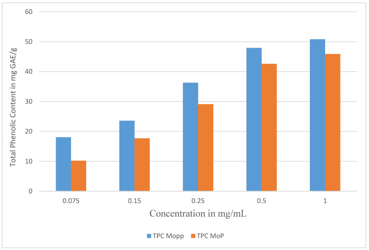
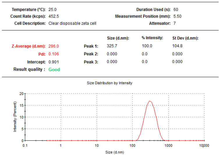
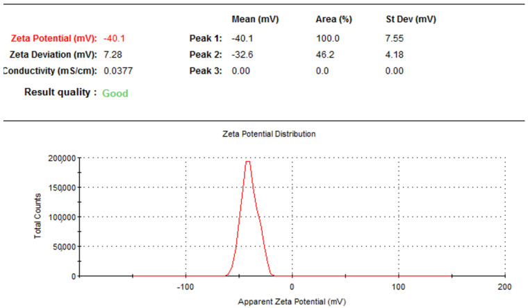
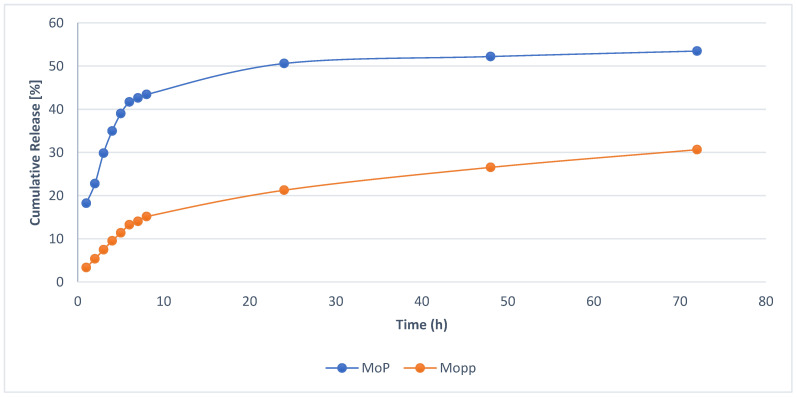
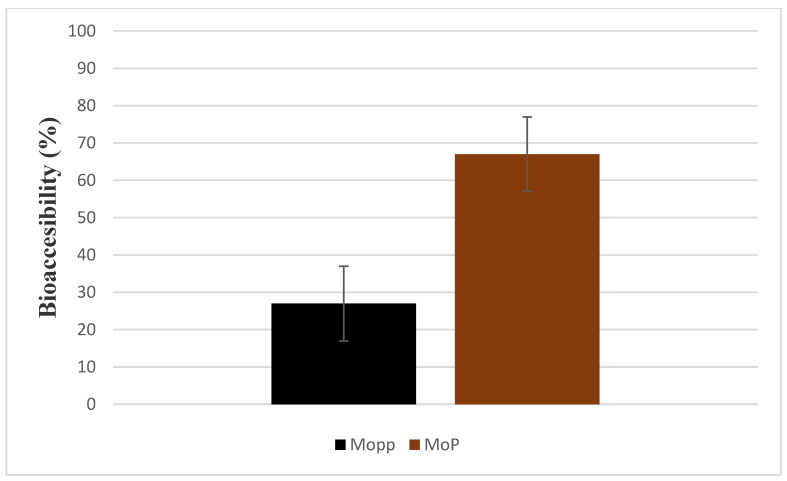
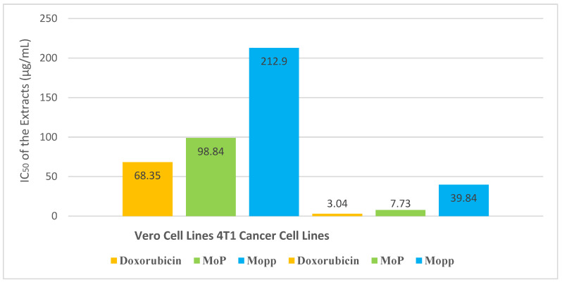
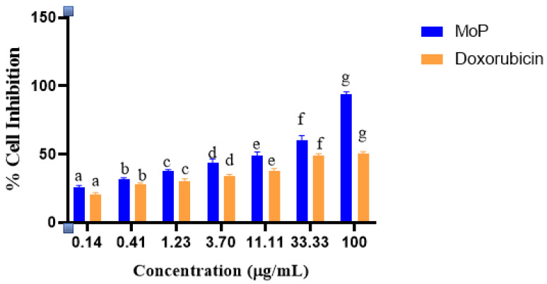
Moringa oleifera leaf polyphenols (Mopp) were encapsulated with phytosomes to enhance their efficacy on 4T1 cancer cell lines. The Mopp were extracted via microwave-assisted extraction. Moringa oleifera polyphenol-loaded phytosomes (MoP) were prepared with the nanoprecipitation method and characterized using the dynamic light scattering and dialysis membrane techniques. The in vitro cytotoxic and antiproliferative activity were investigated with the (3-[4,5-dimethylthiazol-2-yl]-2,5-diphenyltetrazole) MTT assay. Acute toxicity was assessed using Swiss albino mice. An MoP particle size of 296 ± 0.29 nm, −40.1 ± 1.19 mV zeta potential, and polydispersity index of 0.106 ± 0.002 were obtained. The total phenolic content was 50.81 ± 0.02 mg GAE/g, while encapsulation efficiency was 90.32 ± 0.11%. The drug release profiles demonstrated biphasic and prolonged subsequent sustained release. In vitro assays indicated MoP had a low cytotoxicity effect of 98.84 ± 0.53 μg/mL, doxorubicin was 68.35 ± 3.508, and Mopp was 212.9 ± 1.30 μg/mL. Moreover, MoP exhibited the highest antiproliferative effect on 4T1 cancer cells with an inhibitory concentration of 7.73 ± 2.87 μg/mL and selectivity index > 3. The results indicated a significant difference (p ≤ 0.001) in MoP when compared to Mopp and doxorubicin. The in vivo investigation showed the safety of MoP at a dose below 2000 mg/kg. The present findings suggest that MoP may serve as an effective and promising formulation for breast cancer drug delivery and therapy.
Keywords: Moringa oleifera; antiproliferation; breast cancer; natural nanoparticles; polyphenols.
Conflict of interest statement
The authors declare no conflict of interests.
Figures
References
-
- Feng Y., Spezia M., Huang S., Yuan C., Zeng Z., Zhang L., Ji X., Liu W., Huang B., Luo W., et al. Breast cancer development and progression: Risk factors, cancer stem cells, signaling pathways, genomics, and molecular pathogenesis. Genes Dis. 2018;5:77–106. doi: 10.1016/j.gendis.2018.05.001. - DOI - PMC - PubMed
-
- McCormack V., McKenzie F., Foerster M., Zietsman A., Galukande M., Adisa C., Anele A., Parham G., Pinder L.F., Cubasch H., et al. Breast cancer survival and survival gap apportionment in sub-Saharan Africa (ABC-DO): A prospective cohort study. Lancet Glob. Health. 2020;8:1203–1212. doi: 10.1016/S2214-109X(20)30261-8. - DOI - PMC - PubMed
-
- Sood R., Masalu N., Connolly R.M., Chao C.A., Faustine L., Mbulwa C., Anderson B.O., Rositch A.F. Invasive breast cancer treatment in Tanzania: Landscape assessment to prepare for implementation of standardized treatment guidelines. BMC Cancer. 2021;21:527. doi: 10.1186/s12885-021-08252-2. - DOI - PMC - PubMed
MeSH terms
Substances
LinkOut - more resources
Full Text Sources
Medical
