Review
. 2022 Jul 14;27(14):4504.
doi: 10.3390/molecules27144504.

Advances in Fibrin-Based Materials in Wound Repair: A Review

Affiliations
Review

Advances in Fibrin-Based Materials in Wound Repair: A Review

Ilker S Bayer. Molecules. .

Abstract

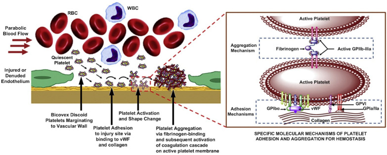
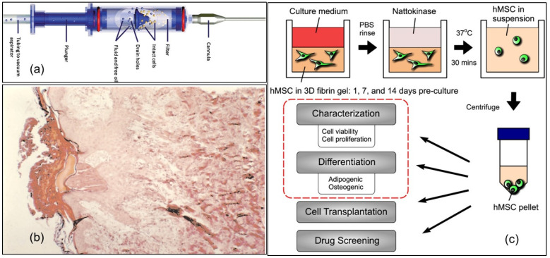
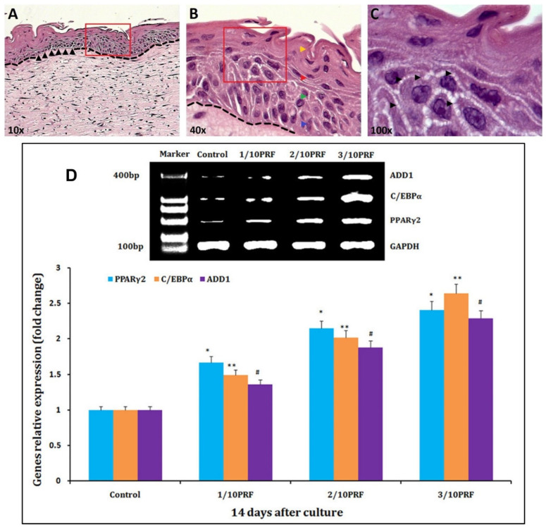
The first bioprocess that occurs in response to wounding is the deterrence of local hemorrhage. This is accomplished by platelet aggregation and initiation of the hemostasis cascade. The resulting blood clot immediately enables the cessation of bleeding and then functions as a provisional matrix for wound healing, which begins a few days after injury. Here, fibrinogen and fibrin fibers are the key players, because they literally serve as scaffolds for tissue regeneration and promote the migration of cells, as well as the ingrowth of tissues. Fibrin is also an important modulator of healing and a host defense system against microbes that effectively maintains incoming leukocytes and acts as reservoir for growth factors. This review presents recent advances in the understanding and applications of fibrin and fibrin-fiber-incorporated biomedical materials applied to wound healing and subsequent tissue repair. It also discusses how fibrin-based materials function through several wound healing stages including physical barrier formation, the entrapment of bacteria, drug and cell delivery, and eventual degradation. Pure fibrin is not mechanically strong and stable enough to act as a singular wound repair material. To alleviate this problem, this paper will demonstrate recent advances in the modification of fibrin with next-generation materials exhibiting enhanced stability and medical efficacy, along with a detailed look at the mechanical properties of fibrin and fibrin-laden materials. Specifically, fibrin-based nanocomposites and their role in wound repair, sustained drug release, cell delivery to wound sites, skin reconstruction, and biomedical applications of drug-loaded fibrin-based materials will be demonstrated and discussed.

Keywords: drug release; fibrin; fibrinogen; nanofibers; protein; wound healing.


Conflict of interest statement

The author declares no conflict of interest.

Figures

Figure 1
Figure 1
(a) Fibrinogen structure. Aα chains are shown in blue, Bβ chains are shown in green, and γ chains are shown in red. Disulfide bridges stabilizing the coiled-coil regions are shown in yellow. Reprinted/adapted with permission from Ref. [5], 2017, DovePress. (b) SEM image of fibrin clots formed using high and low thrombin concentrations after 10 min of lysis after the addition of tissue-type plasminogen activator (tPA) to the surface of the clot. Reprinted/adapted with permission from Ref. [6], 2011, American Society of Hematology. (c) Image of how fibrin fibers from fibrinogen act as glue and a scaffold for the grouping of red blood cells, platelets, and other plasma proteins within a fibrin clot. Reprinted/adapted with permission from Ref. [7], 2017, Elsevier. (d) TEM image (90,000×) of the cross-section of a fibrin fiber. Reprinted/adapted with permission from Ref. [8], 2004, Elsevier.
Figure 2
Figure 2
(a) Fibrinogen to fibrin formation. The α-chain termini fold back on the coiled-coil and interact with the E-region. Fibrin formation involves the cleavage of FpA (orange) by thrombin (fibrin I), which polymerizes into protofibrils. Subsequently, FpB (green) is cleaved by thrombin (fibrin II). FpB cleavage is associated with release of the α-domains, which interact for lateral aggregation. Reprinted/adapted with permission from Ref. [9], 2011, American Heart Association, Inc. (b) The applications of fibrin-based biomaterials are very diverse and mirror the biological roles of fibrin in wound repair. Fibrin-based biomaterials can be applied as a wound healing scaffold to promote hemostasis immediately following injury, and then provide the structure for subsequent inflammatory cell infiltration, angiogenesis, and long-term tissue remodeling. Reprinted/adapted with permission from Ref. [10], 2018, Elsevier.
Figure 3
Figure 3
(a) Results of mechanical testing when the fibrinogen concentration of the glue is varied, keeping the thrombin (200 units/mL), aprotinin (3000 Klu/mL), and calcium (40 μmol/mL) levels constant. (b) Results of mechanical testing when the thrombin concentration of the glue is regulated, keeping the fibrinogen (39 g/L), aprotinin (3000 Klu/mL), and calcium (40 μmol/mL) levels constant. (c) Results of mechanical testing when the factor XIII/fibrinogen ratio is varied, keeping the thrombin (200 units/mL), aprotinin (3000 KIu/mL) and calcium (40 μmol/mL) levels constant. All measurements reported in the plots are the mean p < 0.01, difference from control; Mann–Whitney U test. Data were compiled from [23].
Figure 4
Figure 4
(A) Schematic of the atomic force microscope (AFM) sitting on top of the inverted optical microscope. (B) Top view of the stretched fiber. The initial and stretched states are in dotted gray and solid black, respectively. (C) Typical fibrin fiber stress–strain curve. (DF) Fluorescence microscopy film stills of a stretching experiment. The fiber is anchored on two ridges (brighter, horizontal, 8 μm wide bars) and suspended over a groove (darker, horizontal, 12 μm wide bars); the AFM cantilever appears as a 35 μm wide, dark rectangle; the AFM tip is indicated as a green dot. Reprinted/adapted with permission from Ref. [19], 2010, International Society on Thrombosis and Haemostasis. (G–I) TEM images of negatively contrasted fibrin fibers showing the substructure of branch points. Most branch points consist of three fiber segments of nearly equal diameters that join at a small acute angle with band patterns aligned. The band pattern with a repeat of 22.5 nm is characteristic of fibrin. Scale bar is 0.2 microns. Reprinted/adapted with permission from Ref. [26], 2004, Elsevier.
Figure 5
Figure 5
Physical and biological events in the hemostatic action of platelets. Reprinted/adapted with permission from Ref. [35], 2013, Elsevier.
Figure 6
Figure 6
Schematic description of wound dressings fabricated from sodium carboxymethylcellulose combined with fibrin and seeded with dermal fibroblasts in vitro. (a) Series of porous structures of pure cellulose, cellulose coated with fibrin and cellulose coated with fibrin nanofibers with no cells. (b) Same as (a), but with cell seeding. (c) Images related to the angiogenic potential of various wound dressings assessed with the CAM assay. (ac) Reprinted/adapted with permission from Ref. [48], 2018, MDPI. (d) CAM blood vessel density in different groups. (e) Histological images of CAMs associated with different studied groups stained with Masson’s trichrome (cell nuclei are red and collagen bundles are blue). Statistical significances are shown as * p < 0.05, *** p < 0.001 and **** p < 0.0001 in (d). Reprinted/adapted with permission from Ref. [48], 2022, Elsevier.
Figure 7
Figure 7
(A) Surgery process of canine traumatic hemisection T12 SCI model. (A) Diagram of AFG and (B) the SCI surgery and AFG transplantation process. Reprinted/adapted with permission from Ref. [53], 2022, Oxford University Press. (B) Schematic diagram of the axonal regrowth across the lesion site along the aligned nanofibers. Reproduced with permission from [52]. (C) Photomicrograph of electrospinning aligned fibrin hydrogel. Reprinted/adapted with permission from Ref. [54], 2020, Springer Nature.
Figure 8
Figure 8
(a) Experimental and clinical delivery applications of fibrin in wound healing. Fibrin has been used to deliver cells, drugs, growth factors or gene vectors, and combinations thereof. GF: growth factor, Fbg: fibrinogen, FXIII: factor XIII, MSCs: mesenchymal stem cells, ESC: embryonic stem cell, iPSC: induced pluripotent stem cell, FGF: fibroblast growth factor, PDGF: platelet-derived growth factor, VEGF: vascular endothelial growth factor, IGF: insulin-like growth factor, EGF: epidermal growth factor, KGF: keratinocyte growth factor, NGF: nerve growth factor, BDNF: brain-derived neurotrophic factor, GDNF: glial-cell-line-derived growth factor, NT: neurotrophin, BMP: bone morphogenetic protein, GAM: gene-activated matrix, COPROG: copolymer protected gene vector. Reprinted/adapted with permission from Ref. [62], 2000, Elsevier. (b) Fibrin preparations useful in therapeutic delivery. Reprinted/adapted with permission from Ref. [60], 2001, Elsevier.
Figure 9
Figure 9
(a) Schematic of a modified syringe-based device. The device was designed to harvest small volumes of autologous adipose tissue for cosmetic surgical applications. It aspirates fat at low vacuum into a sterile chamber. Once inside, the fat cells are concentrated by filtration. Fluids and free oil are drawn into a waste canister. Reprinted/adapted with permission from Ref. [73], 2013, Elsevier. (b) Biopsy taken 6 days following the application of fibrin glue suspended keratinocytes combined with allogeneic overgrafting. Histology demonstrates the initial integration of allogeneic dermal elements in the reconstituted neoskin. Reprinted/adapted with permission from Ref. [75], 2014, Mary Ann Liebert, Inc. (c) Illustration depicting the methods by which MSCs were extracted from 3D fibrin gels. Reprinted/adapted with permission from Ref. [76], 2004, Springer Nature.
Figure 10
Figure 10
Histological features of skin organotypic cultures (ORGs) determined by H&E, after 21 days. (A) Whole structure of the ORGs (10×). A clear dermal–epidermal separation (dashed line) and a possible basal membrane (black head arrows) can be seen. (B) The enhanced area (40×) indicated by the red inset in (A). Four differentiation stages of the epidermis: the basal layer (blue head arrow), the spinous layer (green head arrow), the granular layer (red head arrow), and the horny layer (yellow head arrow), and the morphological associated changes in keratinocytes through the different layers of the epidermis. (C) Enhanced (100×) image indicated by the red inset in (B). The black arrows indicate hemidesmosome-like structures between keratinocytes. (AC) Reprinted/adapted with permission from Ref. [97], 2016, Elsevier. (D) mRNA levels of PPARγ2, C/EBPα, and ADD1 mRNA, which are adipogenic marker genes, are higher in the platelet-rich fibrin (PRF) groups than in the control group after 14 days of culture. * p < 0.01, ** p < 0.01, # p < 0.01. Reprinted/adapted with permission from Ref. [98], 2017, Impact Journals, LLC.
Figure 11
Figure 11
Cross-section fibrin fiber models and their corresponding stretch modulus. (A) A fiber with uniformly connected protofibrils has a stretch modulus that is independent of diameter, D. (B) A fiber with a bicycle-spokes-like cross-section can have a stretch modulus that decreases as D−1. (C) Based on experimental observations, however, the stretch modulus scales as D−1.6. This may indicate that the density of connected protofibrils will decrease with increasing fibrin diameter, D, as D−1.6. Reprinted/adapted with permission from Ref. [160], 2021, Elsevier.

References

    1. Janmey P.A., Winer J.P., Weisel J.W. Fibrin gels and their clinical and bioengineering applications. J. R. Soc. Interface. 2009;6:1–10. doi: 10.1098/rsif.2008.0327. - DOI - PMC - PubMed
    1. Doolittle R.F. Fibrinogen and fibrin. Annu. Rev. Biochem. 1984;53:195–229. doi: 10.1146/annurev.bi.53.070184.001211. - DOI - PubMed
    1. Scott E.M., Ariëns R.A., Grant P.J. Genetic and environmental determinants of fibrin structure and function: Relevance to clinical disease. Arterioscler. Thromb. Vasc. Biol. 2004;24:1558–1566. doi: 10.1161/01.ATV.0000136649.83297.bf. - DOI - PubMed
    1. Mosesson M.W. Fibrinogen and fibrin structure and functions. J. Thromb. Haemost. 2005;3:1894–1904. doi: 10.1111/j.1538-7836.2005.01365.x. - DOI - PubMed
    1. Noori A., Ashrafi S.J., Vaez-Ghaemi R., Hatamian-Zaremi A., Webster T.J. A review of fibrin and fibrin composites for bone tissue engineering. Int. J. Nanomed. 2017;12:4937. doi: 10.2147/IJN.S124671. - DOI - PMC - PubMed

LinkOut - more resources