. 2022 Jul 21;27(14):4664.
doi: 10.3390/molecules27144664.

In Silico Target Identification of Galangin, as an Herbal Flavonoid against Cholangiocarcinoma

Affiliations

In Silico Target Identification of Galangin, as an Herbal Flavonoid against Cholangiocarcinoma

Brinda Balasubramanian et al. Molecules. .

Abstract

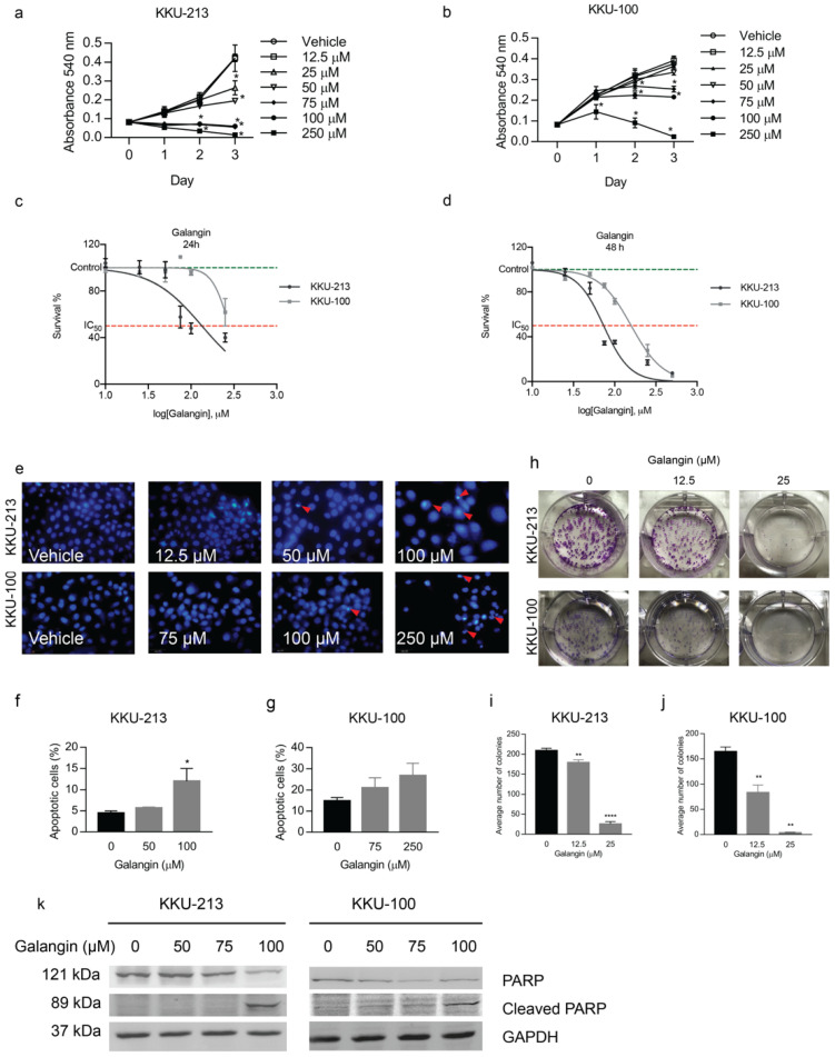
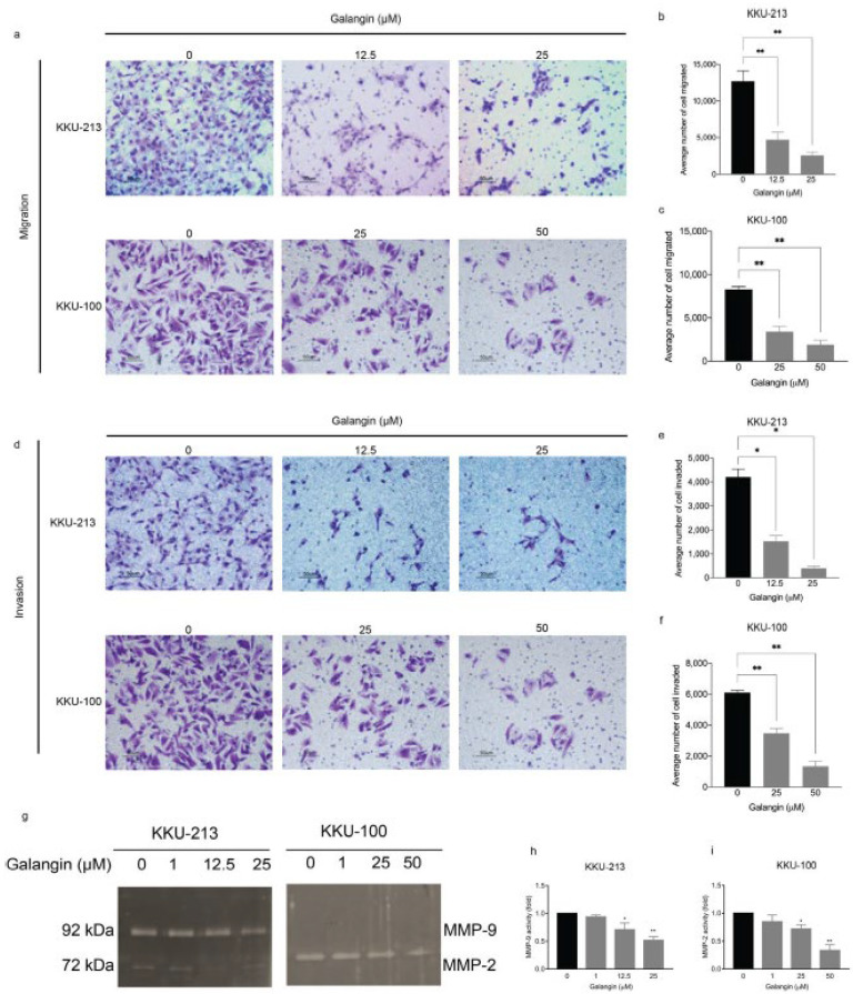
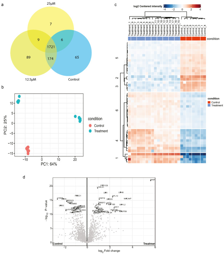
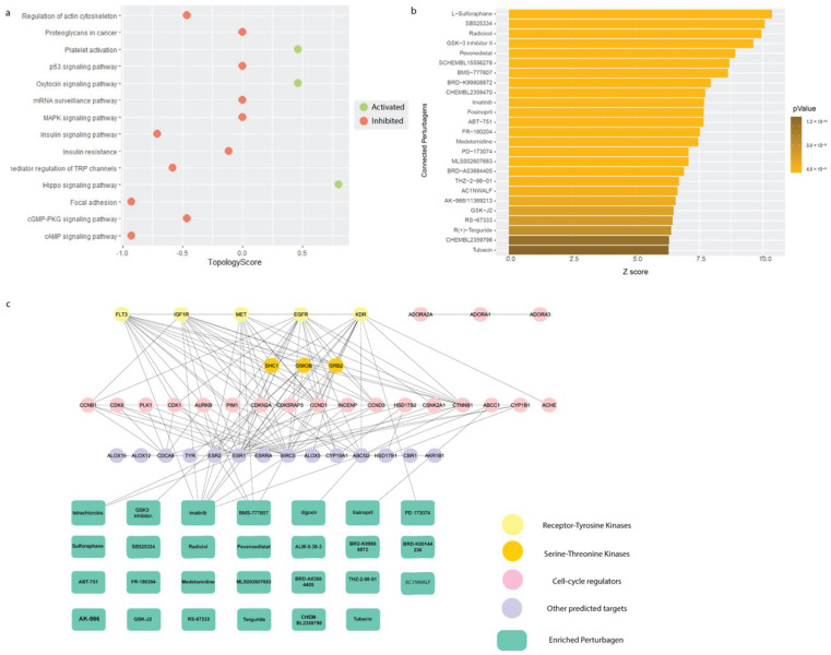
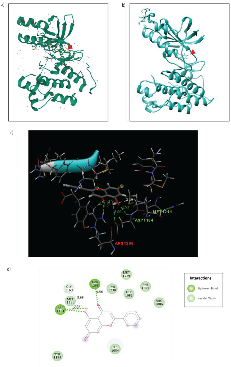
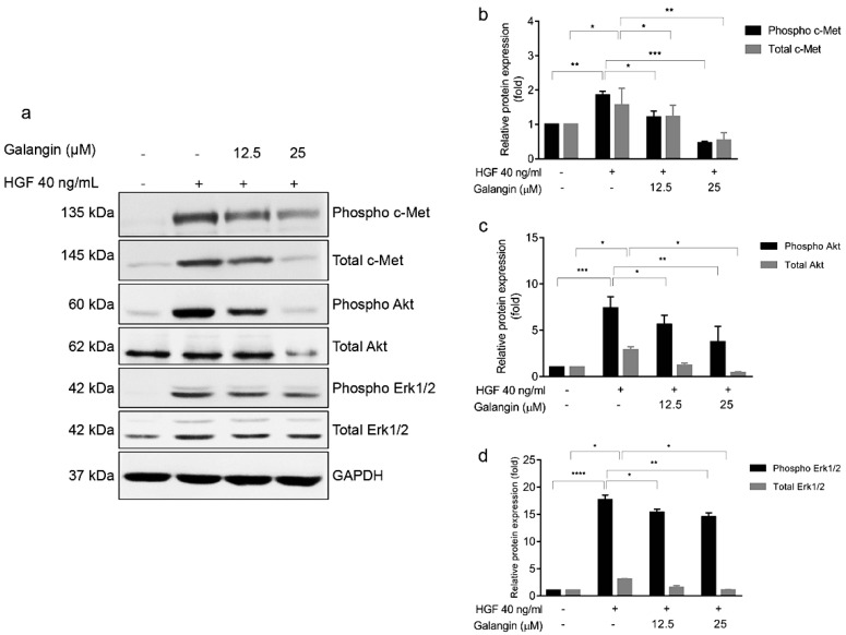
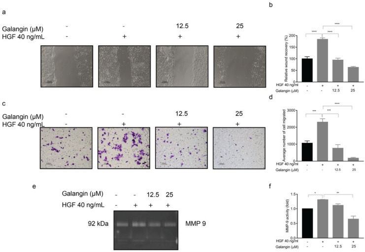
Cholangiocarcinoma (CCA) is a heterogenous group of malignancies in the bile duct, which proliferates aggressively. CCA is highly prevalent in Northeastern Thailand wherein it is associated with liver fluke infection, or Opisthorchis viverrini (OV). Most patients are diagnosed in advanced stages, when the cancer has metastasized or severely progressed, thereby limiting treatment options. Several studies investigate the effect of traditional Thai medicinal plants that may be potential therapeutic options in combating CCA. Galangin is one such herbal flavonoid that has medicinal properties and exhibits anti-tumor properties in various cancers. In this study, we investigate the role of Galangin in inhibiting cell proliferation, invasion, and migration in OV-infected CCA cell lines. We discovered that Galangin reduced cell viability and colony formation by inducing apoptosis in CCA cell lines in a dose-dependent manner. Further, Galangin also effectively inhibited invasion and migration in OV-infected CCA cells by reduction of MMP2 and MMP9 enzymatic activity. Additionally, using proteomics, we identified proteins affected post-treatment with Galangin. Enrichment analysis revealed that several kinase pathways were affected by Galangin, and the signature corroborated with that of small molecule kinase inhibitors. Hence, we identified putative targets of Galangin using an in silico approach which highlighted c-Met as candidate target. Galangin effectively inhibited c-Met phosphorylation and subsequent signaling in in vitro CCA cells. In addition, Galangin was able to inhibit HGF, a mediator of c-Met signaling, by suppressing HGF-stimulated invasion, as well as migration and MMP9 activity. This shows that Galangin can be a useful anti-metastatic therapeutic strategy in a subtype of CCA patients.

Keywords: Galangin; anti-cancer; cholangiocarcinoma; molecular docking; proteomics.


Conflict of interest statement

The authors declare no conflict of interest.

Figures

Figure 1
Figure 1
Effect of Galangin on cholangiocarcinoma cell proliferation. The cytotoxic effect of Galangin on (a) KKU-213, (b) KKU-100, and CCA cell lines were determined by MTT assay. The IC50 values were calculated using the dose-response curve for (c) 24 h and (d) 48 h of treatment. (e) Detection of apoptosis by DAPI staining after Galangin treatment in CCA cell lines in various concentrations. The number of apoptotic cells were quantified in (f) KKU-213 and (g) KKU-100 cell lines through FACS analysis. (h) Representative images of colony formation and quantification for average number of colonies on (i) KKU-213 and (j) KKU-100 cells upon Galangin treatment at 10 and 25 µM. (k) Effect of Galangin on protein involved in apoptosis. Cellular proteins were analyzed by Western blot assay for the expression of PARP and cleaved PARP. Values represents the mean ± SEM of three independent experiments, * p ≤ 0.05, ** p ≤ 0.01, **** p ≤ 0.0001 compared with the untreated control.
Figure 2
Figure 2
Effect of Galangin on migration and invasion in KKU-213 and KKU-100 CCA cells. Transwell assay was used to detect cell migration and invasion. Images were taken at 20× magnification. Images of (a) cell migration, each cell group treated with various of Galangin, and their respective quantifications in (b) KKU-213 and (c) KKU-100 cells. Images of (d) cell invasion showing each cell group treated with various amounts of Galangin, and their respective quantifications in (e) KKU-213 and (f) KKU-100 cells. (g) Representative images of the gelatin zymography and (h) quantification the band intensity of MMP-9 activity in KKU-213 and (i) quantification of the band intensity of MMP-2 activity in KKU-100 after Galangin treatment in various concentrations. * p ≤ 0.05, ** p ≤ 0.01.
Figure 3
Figure 3
Proteomics analysis upon Galangin treatment in KKU-213 cells. (a) Venn Diagram of uniquely identified peptides in each condition. (b) Principle-component analysis of control and Galangin treated samples. (c) Heatmap of differentially expressed proteins between treatment and control with the significance criteria of proteomics fold change at log2 (1.5) and p < 0.05. (d) Volcano plot depicting the differentially expressed proteins (DEP) between control and treatment samples.
Figure 4
Figure 4
Pathway enrichment and pharmaco-connectivity analysis of the DEP signature. (a) Signaling pathway impact analysis (SPIA) of the DEP signature. Green nodes represent pathways that are activated (topology score > 0), and red nodes represent pathways that are inhibited (topology score < 0). (b) Significant positively connected perturbagens to the DEP signature, ranked by lowest p-value and highest Z-score. (c) Pharmaco-connectivity analysis between the positively connected perturbagens and the predicted targets of Galangin based on structural similarity.
Figure 5
Figure 5
Molecular docking of Galangin to the tyrosine kinase domain of the c-Met receptor. (a) 3D X-ray crystallography structure of c-Met inhibitor, AM7, bound to c-Met (PDB: 2RFN). (b) 3D Docking model of Galangin (PDB: 57D docked to the c-Met receptor (PDB:1R1W) using SwissDock. (c) 3D interaction plot of Galangin binding to c-Met receptor with distance in (Å). (d) 2D interaction plot of Galangin binding to c-Met receptor. The important interactions were highlighted, including conventional hydrogen bond (dark green dot line) and van der Waals (light green).
Figure 6
Figure 6
Galangin treatment with HGF-stimulated KKU-213 cells affects c-Met signaling. (a) Effect of Galangin on HGF-stimulated cell signaling by western blotting. Cells were analyzed for (b) phospho c-Met, total c-Met, (c) phospho Akt, total Akt, (d) phospho ERK1/2, and total ERK1/2 expression by Western blotting. GAPDH was used as loading control to normalize the amount of protein in each experiment conditions. Data are the mean ± SEM from three independent experiments, * p ≤ 0.05, ** p ≤ 0.01, *** p ≤ 0.001, **** p ≤ 0.0001 compared with the HGF treatment.
Figure 7
Figure 7
Effect of Galangin upon HGF-stimulation on wound-healing, cell migration, and MMP-9 activity in KKU-213 CCA cells. KKU-213 cells were stimulated with HGF and treated with various concentrations of Galangin. Representative images of (a) wound-healing and its (b) quantitation. Representative images of (c) Transwell cell migration and its respective (d) quantitation. Representative images of (e) MMP-9 activity and (f) its quantitation. Data are the mean ± SEM from three independent experiments, * p ≤ 0.05, ** p ≤ 0.01, *** p ≤ 0.001, **** p ≤ 0.0001 compared with the HGF treatment.

References

    1. Sriamporn S., Pisani P., Pipitgool V., Suwanrungruang K., Kamsa-ard S., Parkin D.M. Prevalence of Opisthorchis viverrini infection and incidence of cholangiocarcinoma in Khon Kaen, Northeast Thailand. Trop. Med. Int. Health. 2004;9:588–594. doi: 10.1111/j.1365-3156.2004.01234.x. - DOI - PubMed
    1. Pairojkul C. Liver fluke and cholangiocarcinoma in Thailand. Pathology. 2014;46:S24. doi: 10.1097/01.PAT.0000454133.12330.87. - DOI
    1. Leelawat K., Leelawat S., Tepaksorn P., Rattanasinganchan P., Leungchaweng A., Tohtong R., Sobhon P. Involvement of c-Met/hepatocyte growth factor pathway in cholangiocarcinoma cell invasion and its therapeutic inhibition with small interfering RNA specific for c-Met. J. Surg. Res. 2006;136:78–84. doi: 10.1016/j.jss.2006.05.031. - DOI - PubMed
    1. Ramirez-Merino N., Aix S.P., Cortes-Funes H. Chemotherapy for cholangiocarcinoma: An update. World J. Gastrointest. Oncol. 2013;5:171–176. doi: 10.4251/wjgo.v5.i7.171. - DOI - PMC - PubMed
    1. Leyva-Illades D., McMillin M., Quinn M., Demorrow S. Cholangiocarcinoma pathogenesis: Role of the tumor microenvironment. Transl. Gastrointest. Cancer. 2012;1:71–80. - PMC - PubMed

MeSH terms