Review
. 2022 Jul 20;12(14):2485.
doi: 10.3390/nano12142485.

Biomimetic Nanomaterials: Diversity, Technology, and Biomedical Applications

Affiliations
Review

Biomimetic Nanomaterials: Diversity, Technology, and Biomedical Applications

Kamil G Gareev et al. Nanomaterials (Basel). .

Abstract

Biomimetic nanomaterials (BNMs) are functional materials containing nanoscale components and having structural and technological similarities to natural (biogenic) prototypes. Despite the fact that biomimetic approaches in materials technology have been used since the second half of the 20th century, BNMs are still at the forefront of materials science. This review considered a general classification of such nanomaterials according to the characteristic features of natural analogues that are reproduced in the preparation of BNMs, including biomimetic structure, biomimetic synthesis, and the inclusion of biogenic components. BNMs containing magnetic, metal, or metal oxide organic and ceramic structural elements (including their various combinations) were considered separately. The BNMs under consideration were analyzed according to the declared areas of application, which included tooth and bone reconstruction, magnetic and infrared hyperthermia, chemo- and immunotherapy, the development of new drugs for targeted therapy, antibacterial and anti-inflammatory therapy, and bioimaging. In conclusion, the authors' point of view is given about the prospects for the development of this scientific area associated with the use of native, genetically modified, or completely artificial phospholipid membranes, which allow combining the physicochemical and biological properties of biogenic prototypes with high biocompatibility, economic availability, and scalability of fully synthetic nanomaterials.

Keywords: applications; biomedicine; biomimetics; nanomaterials; nanoparticles; synthesis technique; theranostics.


Conflict of interest statement

The authors declare no conflict of interest.

Figures

Figure 1
Figure 1
Biomimetic nanoparticles for biomedical applications. (a) Technology evolution: Early generations of particles were biologically inert and covered with nonfouling coatings, preventing their nonspecific interactions with the cells they encountered in vivo. From here, the next generation of nanoparticles became active, targeting molecules, which enabled the nanoparticles to reach the disease site and engage with the local environment. Taking inspiration from nature, the third generation of cell-membrane-based biomimetic nanoparticles mimicked the surface features of native cells by utilizing the whole cell membrane or membrane protein functionalization onto synthetic carriers (Reprinted from [31], license CC BY 4.0.) (b) Schematic presentation of different strategies of inflammation-targeting biomimetic nanoparticles. Orange and red spheres represent drug-encapsulated synthetic nanoparticles (grey) and liposomes (green), respectively. (Reprinted from [16], license CC BY 4.0.) (c) An example of modern BNM concept implementation: cell-membrane-coated NPs designed for atherosclerosis and inflammation therapy. The membranes of RBCs, platelets, and macrophages are extracted and used to coat different kinds of NPs depending on the site of inflammation and atherosclerosis. Each cell membrane has its own unique surface proteins, such as CD47 on RBC, integrin a4b1 on macrophages, and GPIIb/IIa on platelets, modifying the therapeutic effects. (Reprinted from [20], license CC BY 4.0.)
Figure 1
Figure 1
Biomimetic nanoparticles for biomedical applications. (a) Technology evolution: Early generations of particles were biologically inert and covered with nonfouling coatings, preventing their nonspecific interactions with the cells they encountered in vivo. From here, the next generation of nanoparticles became active, targeting molecules, which enabled the nanoparticles to reach the disease site and engage with the local environment. Taking inspiration from nature, the third generation of cell-membrane-based biomimetic nanoparticles mimicked the surface features of native cells by utilizing the whole cell membrane or membrane protein functionalization onto synthetic carriers (Reprinted from [31], license CC BY 4.0.) (b) Schematic presentation of different strategies of inflammation-targeting biomimetic nanoparticles. Orange and red spheres represent drug-encapsulated synthetic nanoparticles (grey) and liposomes (green), respectively. (Reprinted from [16], license CC BY 4.0.) (c) An example of modern BNM concept implementation: cell-membrane-coated NPs designed for atherosclerosis and inflammation therapy. The membranes of RBCs, platelets, and macrophages are extracted and used to coat different kinds of NPs depending on the site of inflammation and atherosclerosis. Each cell membrane has its own unique surface proteins, such as CD47 on RBC, integrin a4b1 on macrophages, and GPIIb/IIa on platelets, modifying the therapeutic effects. (Reprinted from [20], license CC BY 4.0.)
Figure 2
Figure 2
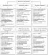
Classification of biomimetic nanomaterials (BNMs) based on the literature data, including information on biomimetic structure [16,34,35,36,37,38], biomimetic synthesis [32,39,40,41,42,43], biogenic components [31,33,44,45,46,47], magnetic BNMs [48,49,50,51,52,53,54], metal and metal oxide BNMs [55,56,57,58,59,60,61], organic, ceramic and hybrid BNMs [62,63,64,65,66,67].
Figure 3
Figure 3
Chitosan-oligosaccharide-coated biocompatible palladium nanoparticles (Pd@COS NPs) for photo-based imaging and therapy. (a) A scheme showing the preparation of Pd NPs, further surface coating with thiolated chitosan oligosaccharide (Pd@COS NPs), and finally, functionalization using an RGD peptide (Pd@COS-RGD). (b) A systematic illustration showing the photothermal ablation and photoacoustic imaging of tumor tissue using Pd@COS-RGD. (Reprinted from [69], license CC BY 4.0.)
Figure 4
Figure 4
Schematic illustration of encapsulation procedure and release mechanism of biocompatible upconversion nanoparticles (UCNP). (a) Encapsulation of UCNPs in a novel, synthesized phosphate surfactant through sonication at rt. (b) Release of UCNPs after a specific cleavage of phosphate surfactant by the sPLA-2 enzyme. (Reprinted from [75], license CC BY 4.0.)
Figure 5
Figure 5
The four main routes of the cytotoxic mechanism of AgNPs. 1: AgNPs adhere to the surface of a cell, damaging its membrane and altering the transport activity; 2: AgNPs and Ag ions penetrate inside the cell and interact with numerous cellular organelles and biomolecules, which can affect corresponding cellular functions; 3: AgNPs and Ag ions participate in the generation of reactive oxygen species (ROS) inside the cell, leading to cell damage; and 4: AgNPs and Ag ions induce the genotoxicity. (Reprinted from [81], license CC BY 4.0.)
Figure 6
Figure 6
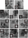
HRTEM images of different NPs: (a,b) inorganic magnetite NPs, (ce) MamC magnetite NPs, (fh) Mms6 magnetite NPs, and (ik) Mms6-MamC-mediated NPs. Selected areas of electron diffraction are shown for each sample. (Reprinted from [32], license CC BY 4.0.)
Figure 7
Figure 7
Magnetofection for gene delivery: (A) schematic representation of the process and (B) schematic illustration of DNA loading into lamellar magnetic hydroxyapatite (MHAp) nanoparticles for nucleic acid delivery. (Reprinted from [35], license CC BY 3.0.)
Figure 8
Figure 8
Surface features and scanning electron micrographs of a TiUnite dental implant surface. (Reprinted from [38], license CC BY 4.0.)
Figure 9
Figure 9
Schematic representation of the mechanism and final outcomes of the interaction of Au NPs and Ag NPs with a water dispersion of cubosomes and solid-supported films of cubosomes. (Reprinted from [34], license CC BY 4.0.)
Figure 10
Figure 10
Schematic illustration of biomimetically mineralized metal–organic framework (MOF). (a) Schematic of a sea urchin, a hard, porous, protective shell that is biomineralized by soft biological tissue. (b) Schematic of an MOF biocomposite showing a biomacromolecule (for example, protein, enzyme, or DNA) encapsulated within a porous, crystalline shell. (Reprinted from [108], license CC BY 4.0.)
Figure 11
Figure 11
Schematic illustration of mechanism of mitochondria-targeted cancer cell membrane biomimetic metal–organic framework mediated sonodynamic therapy and immune checkpoint blockade immunotherapy. (Reprinted from [115], license CC BY 4.0.)
Figure 12
Figure 12
Schematic of anisotropic nanoparticle fabrication and RBC membrane coating. (A) Spherical PLGA nanoparticles (NPs) were synthesized and cast onto a thin plastic film of 10% polyvinyl alcohol (PVA) and 2% glycerol. Particles were then stretched under heat in one and two dimensions (2D) to generate prolate and oblate ellipsoidal particles, respectively. (B) RBCs underwent hypotonic lysis and were then sonicated to generate sub—200 nm vesicles. RBC-derived vesicles were then coated on PLGA nanoparticles of all shapes under sonication. (Reprinted from [33], license CC BY 4.0.)
Figure 13
Figure 13
Plausible mechanism for the formation of Ag–TiO2 NCs using Origanum majorana leaf extract. (Reprinted from [40], license CC BY 4.0).
Figure 14
Figure 14
Main applications of biomimetic nanomaterials.

References

    1. Zan G., Wu Q. Biomimetic and Bioinspired Synthesis of Nanomaterials/Nanostructures. Adv. Mater. 2016;28:2099–2147. doi: 10.1002/adma.201503215. - DOI - PubMed
    1. Sheikhpour M., Barani L., Kasaeian A. Biomimetics in drug delivery systems: A critical review. J. Control. Release. 2017;253:97–109. doi: 10.1016/j.jconrel.2017.03.026. - DOI - PubMed
    1. Das D., Noh I. Biomimetic Medical Materials: From Nanotechnology to 3D Bioprinting. Springer; Berlin/Heidelberg, Germany: 2018.
    1. Ruys A.J. Biomimetic Biomaterials. Elsevier; Amsterdam, The Netherlands: 2013.
    1. Poupon E., Nay B. Biomimetic Organic Synthesis. Volume 1–2. Wiley; Hoboken, NJ, USA: 2011.

LinkOut - more resources