. 2022 Jul 14;14(14):2894.
doi: 10.3390/nu14142894.

β-Sitosterol Attenuates Dexamethasone-Induced Muscle Atrophy via Regulating FoxO1-Dependent Signaling in C2C12 Cell and Mice Model

Affiliations

β-Sitosterol Attenuates Dexamethasone-Induced Muscle Atrophy via Regulating FoxO1-Dependent Signaling in C2C12 Cell and Mice Model

Young-Sool Hah et al. Nutrients. .

Abstract

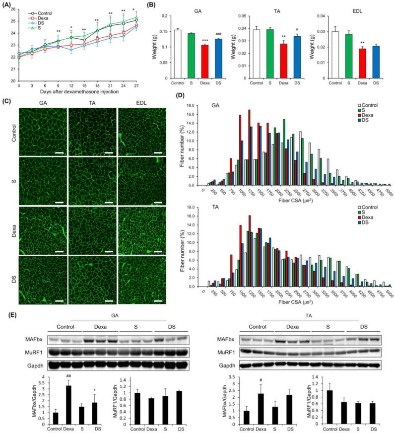
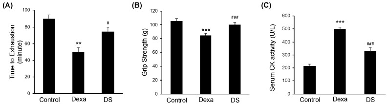
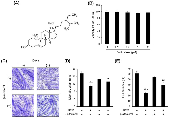
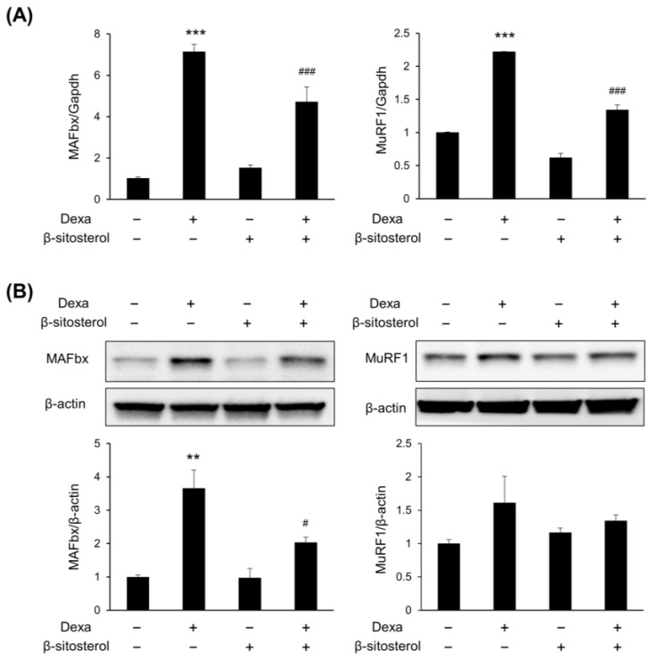
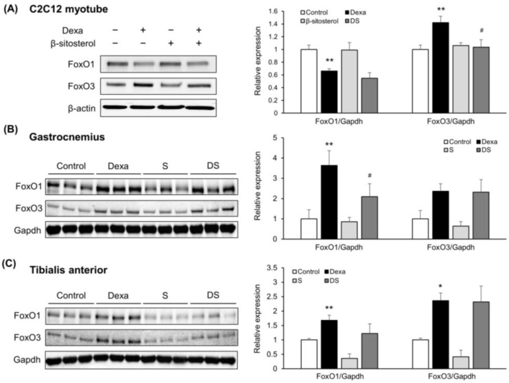
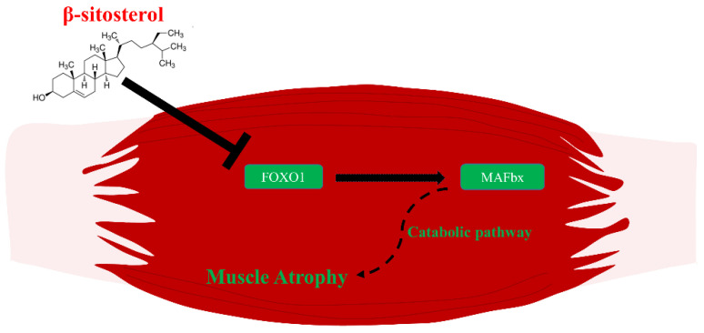
Sarcopenia refers to a decline in muscle mass and strength with age, causing significant impairment in the ability to carry out normal daily functions and increased risk of falls and fractures, eventually leading to loss of independence. Maintaining protein homeostasis is an important factor in preventing muscle loss, and the decrease in muscle mass is caused by an imbalance between anabolism and catabolism of muscle proteins. Although β-sitosterol has various effects such as anti-inflammatory, protective effect against nonalcoholic fatty liver disease (NAFLD), antioxidant, and antidiabetic activity, the mechanism of β-sitosterol effect on the catabolic pathway was not well known. β-sitosterol was assessed in vitro and in vivo using a dexamethasone-induced muscle atrophy mice model and C2C12 myoblasts. β-sitosterol protected mice from dexamethasone-induced muscle mass loss. The thickness of gastrocnemius muscle myofibers was increased in dexamethasone with the β-sitosterol treatment group (DS). Grip strength and creatine kinase (CK) activity were also recovered when β-sitosterol was treated. The muscle loss inhibitory efficacy of β-sitosterol in dexamethasone-induced muscle atrophy in C2C12 myotube was also verified in C2C12 myoblast. β-sitosterol also recovered the width of myotubes. The protein expression of muscle atrophy F-box (MAFbx) was increased in dexamethasone-treated animal models and C2C12 myoblast, but it was reduced when β-sitosterol was treated. MuRF1 also showed similar results to MAFbx in the mRNA level of C2C12 myotubes. In addition, in the gastrocnemius and tibialis anterior muscles of mouse models, Forkhead Box O1 (FoxO1) protein was increased in the dexamethasone-treated group (Dexa) compared with the control group and reduced in the DS group. Therefore, β-sitosterol would be a potential treatment agent for aging sarcopenia.

Keywords: FoxO1; MAFbx; MuRF1; dexamethasone; muscle atrophy; β-sitosterol.


Conflict of interest statement

The authors declare no conflict of interest.

Figures

Figure 1
Figure 1
β-sitosterol prevents Dexa-induced muscle atrophy in mice. (A) Body weight. * p < 0.05 and ** p < 0.01 control vs. Dexa. (B) Muscle weight of GA, TA, and EDL. ** p < 0.01 and *** p < 0.001 vs. control. # p < 0.05 and ### p < 0.001 vs. Dexa. (C) Representative immunofluorescent staining of myofiber cross section of GA, TA, and EDL. A microscope with a 10× objective was used to capture the images. The scale bar represents 100 μm. (D) Quantification of myofiber size by cross-sectional area (CSA) measurements for GA and TA muscle. Data are shown as mean ± S.E. (n = 9 per group). (E) Representative images of the western blot analyses for MAFbx and MuRF1 in GA and TA. # p < 0.05, ## p < 0.01 vs. control; * p < 0.05 vs. Dexa. GA—gastrocnemius muscles; TA—tibialis anterior; EDL—extensor digitorum longus; Dexa—Dexamethasone; S—β-sitosterol; DS—dexa + β-sitosterol.
Figure 2
Figure 2
β-sitosterol ameliorates Dexa-induced muscle dysfunction in mice. (A) Forced running time. (B) Grip strength. (C) Serum CK activity. ** p < 0.01 and *** p < 0.001 compared to the untreated control. # p < 0.05, ### p < 0.001 compared to the to the Dexa treated control. DS—dexa + β-sitosterol; CK—creatine kinase.
Figure 3
Figure 3
β-sitosterol attenuates Dexa-induced muscle atrophy in C2C12 myotubes. (A) Chemical structure of β-sitosterol. (B) C2C12 cells were cultured in medium with various concentrations (0.25, 0.5, 1, and 2 mM) of β-sitosterol for 24 h. Cell viability was measured by a CCK-8 assay. The viability of control (untreated cells) was set to 100%. Viability as a percentage of that of control cells is shown. Bars represent the mean ± S.D. (n = 4 per treatment). (C) May–Grunwald and Giemsa staining. The cells were incubated with 0.5 mM β-sitosterol for 48 h in the presence or absence of 1 μM dexamethasone (Dexa) for 48 h. (D) Quantification of myotube widths from May–Grunwald and Giemsa stained images. Data shown are mean ± S.D. (n = 100). *** p < 0.001 vs. untreated control; ## p < 0.01 vs. Dexa. (E) Fusion index. The total number of nuclei incorporated in myotubes and the total number of nuclei were scored. Fusion index was calculated as the percentage of total nuclei incorporated in myotubes. *** p < 0.001 vs. untreated control, ## p < 0.01 vs. Dexa. DS—dexa + β-sitosterol.
Figure 4
Figure 4
β-sitosterol suppresses the expression of MAFbx and MuRF1. C2C12 myotubes were treated with 0.5 mM β-sitosterol in the presence or absence of 1 μM Dexa for 48 h. (A) MAFbx and MuRF1 mRNA levels were analyzed by quantitative PCR. GAPDH was used as a control. Data are shown as mean ± S.D. (n = 4 per group). *** p < 0.001 vs. control; ### p < 0.001 vs. Dexa. (B) The expression of MAFbx and MuRF1 protein in C2C12 myotubes was estimated by Western blot analysis. β-actin was used as a control for protein loading. ** p < 0.01 vs. control, # p < 0.05 vs. Dexa.
Figure 5
Figure 5
β-sitosterol inhibits Dexa-induced atrophy in C2C12 myotube and mice via FoxO1/3-dependent signaling. (A) Representative images of the Western blot analyses for FoxO1 and FoxO3 in C2C12 myotube. Right panel show quantification of the indicated proteins. Data shown are mean ± S.D. (n = 3 per group). ** p < 0.01 vs. control; # p < 0.05 vs. Dexa. (B) Representative images of the Western blot analyses for FoxO1 and FoxO3 in gastrocnemius muscles. Right panel show quantification of the indicated proteins. Data shown are mean ± S.D. (n = 3 per group). ** p < 0.01 vs. control; # p < 0.05 vs. Dexa. (C) Representative images of the Western blot analyses for FoxO1 and FoxO3 in tibialis anterior muscles. Right panel show quantification of the indicated proteins. Data shown are mean ± S.D. (n = 3 per group). ** p < 0.01; * p < 0.05 vs. control. Dexa—Dexamethasone; S—β-sitosterol; DS—dexa + β-sitosterol.
Figure 6
Figure 6
The overall β-sitosterol mechanisms related to muscle atrophy. β-sitosterol down-regulates the transcriptional factor, FoxO1. Down-regulated FoxO1 becomes unable to affect the expression of MAFbx. Consequently, inhibited MAFbx cannot induce muscle atrophy via the catabolic pathway.

References

    1. Faulkner J.A., Larkin L.M., Claflin D.R., Brooks S.V. Age-related changes in the structure and function of skeletal muscles. Clin. Exp. Pharmacol. Physiol. 2007;34:1091–1096. doi: 10.1111/j.1440-1681.2007.04752.x. - DOI - PubMed
    1. Rosenberg I.H. Sarcopenia: Origins and clinical relevance. J. Nutr. 1997;127((Suppl. 5)):990S–991S. doi: 10.1093/jn/127.5.990S. - DOI - PubMed
    1. Lipovec N.C., Schols A.M., Borst B.V.D., Beijers R.J., Kosten T., Omersa D., Lainscak M. Sarcopenia in Advanced COPD Affects Cardiometabolic Risk Reduction by Short-Term High-intensity Pulmonary Rehabilitation. J. Am. Med. Dir. Assoc. 2016;17:814–820. doi: 10.1016/j.jamda.2016.05.002. - DOI - PubMed
    1. Kang S.-Y., Lim G.E., Kim Y.K., Kim H.W., Lee K., Park T.-J., Kim J. Association between Sarcopenic Obesity and Metabolic Syndrome in Postmenopausal Women: A Cross-sectional Study Based on the Korean National Health and Nutritional Examination Surveys from 2008 to 2011. J. Bone Metab. 2017;24:9–14. doi: 10.11005/jbm.2017.24.1.9. - DOI - PMC - PubMed
    1. Kim J.H., Cho J.J., Park Y.S. Relationship between sarcopenic obesity and cardiovascular disease risk as estimated by the Framingham risk score. J. Korean Med. Sci. 2015;30:264–271. doi: 10.3346/jkms.2015.30.3.264. - DOI - PMC - PubMed