. 2022 Jul 20;14(14):2965.
doi: 10.3390/nu14142965.

Neutrophil Extracellular Traps Promote NLRP3 Inflammasome Activation and Glomerular Endothelial Dysfunction in Diabetic Kidney Disease

Affiliations

Neutrophil Extracellular Traps Promote NLRP3 Inflammasome Activation and Glomerular Endothelial Dysfunction in Diabetic Kidney Disease

Anubhuti Gupta et al. Nutrients. .

Erratum in

Abstract

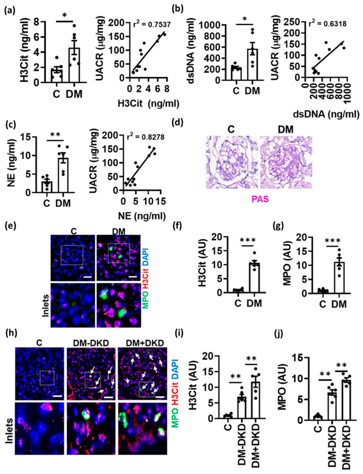
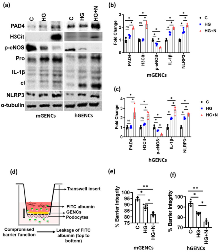
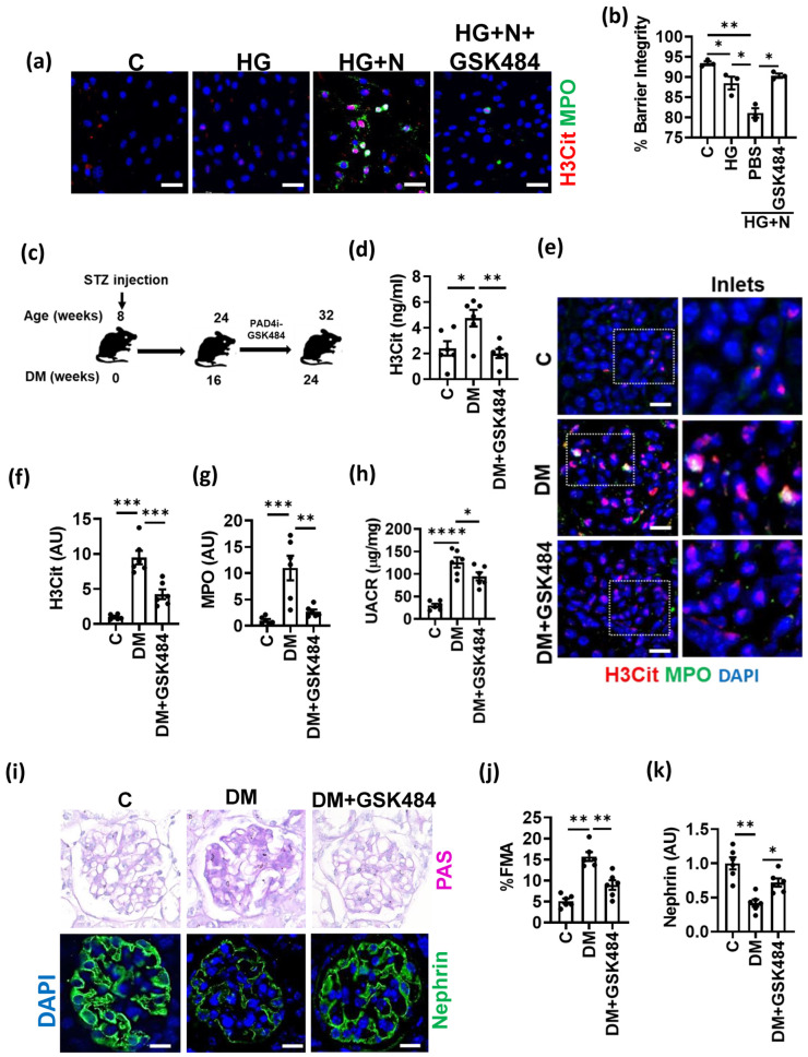
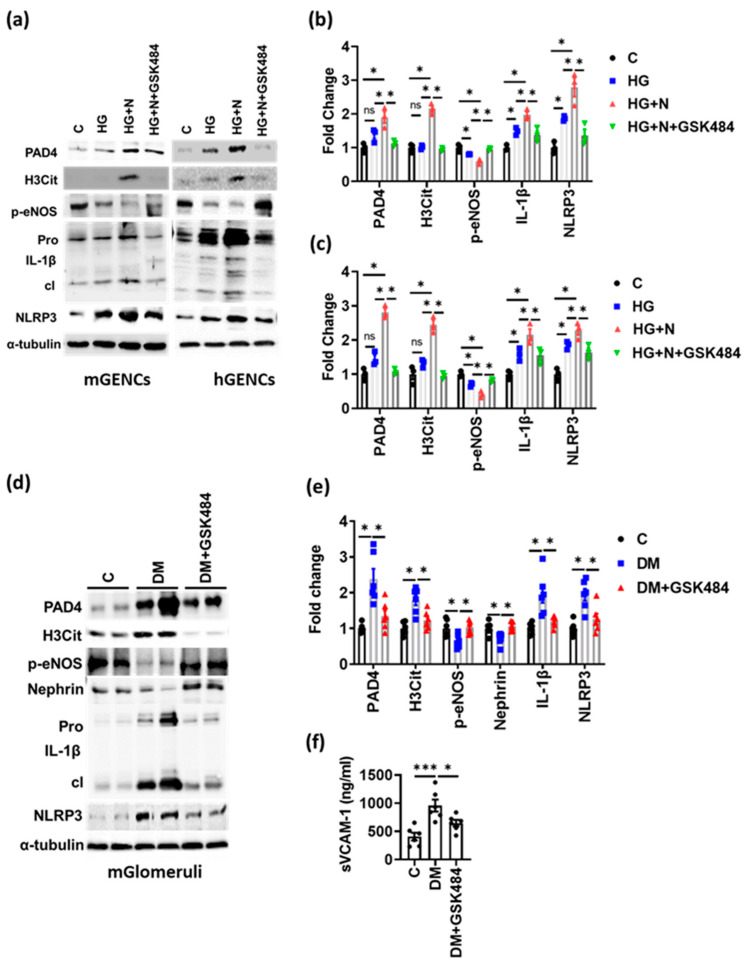
Diabetes mellitus is a metabolic disease largely due to lifestyle and nutritional imbalance, resulting in insulin resistance, hyperglycemia and vascular complications. Diabetic kidney disease (DKD) is a major cause of end-stage renal failure contributing to morbidity and mortality worldwide. Therapeutic options to prevent or reverse DKD progression are limited. Endothelial and glomerular filtration barrier (GFB) dysfunction and sterile inflammation are associated with DKD. Neutrophil extracellular traps (NETs), originally identified as an innate immune mechanism to combat infection, have been implicated in sterile inflammatory responses in non-communicable diseases. However, the contribution of NETs in DKD remains unknown. Here, we show that biomarkers of NETs are increased in diabetic mice and diabetic patients and that these changes correlate with DKD severity. Mechanistically, NETs promote NLRP3 inflammasome activation and glomerular endothelial dysfunction under high glucose stress in vitro and in vivo. Inhibition of NETs (PAD4 inhibitor) ameliorate endothelial dysfunction and renal injury in DKD. Taken together, NET-induced sterile inflammation promotes diabetes-associated endothelial dysfunction, identifying a new pathomechanism contributing to DKD. Inhibition of NETs may be a promising therapeutic strategy in DKD.

Keywords: NLRP3 inflammasome; diabetic kidney disease; endothelial dysfunction; glomerular endothelial cells; glomerular filtration barrier disruption; neutrophil extracellular traps.


Conflict of interest statement

The authors declare no conflict of interest.

Figures

Figure 1
Figure 1
Increased NETs in diabetic kidney disease. (ac) Plasma levels of H3Cit, dsDNA and neutrophil elastase, NE (bar graphs with dot-plot) in non-diabetic control (C) and diabetic (DM, STZ-model) mice. (d) Representative images showing PAS staining in diabetic (DM) versus non-diabetic control (C) mice. (ej) Immunofluorescence staining ((e,h): representative images; (f,g,i,j): bar graphs with dotplot summarizing results) of glomeruli showing H3Cit staining (red) and MPO staining (green) in control (C) and diabetic mice (DM, (eg)) and human kidneys from non-diabetic controls (C) and diabetic patients without (DM-DKD) or with (DM+DKD) diabetic kidney disease (hj). Correlation (Pearson’s correlation) of H3Cit, dsDNA and NE with urinary albumin creatinine ratio (UACR, line graphs, ac). Each dot represents one mouse ((ac,f,g), n = 6 mice each group) or one human sample (i,j); (e,h): scale bar, 50 μm; (ac,f,g,i,j): * p < 0.05, ** p < 0.01, *** p < 0.001, t test.
Figure 2
Figure 2
Neutrophils promote inflammasome activation and endothelial dysfunction. (a,b) Immunoblots ((a), representative blots; (b), bar graph summarizing results) reflecting NET markers (PAD4, H3Cit), inflammasome activation (IL-1β: cleaved form—cl, inactive preform—Pro; NLRP3) and endothelial function (p-eNOS) in mouse (mGENCs) and (c) human (hGENCs) glomerular endothelial cells upon exposure to high glucose and neutrophils (HG+N) compared to high glucose alone (HG, 25 mM) or control (C, PBS). (d) Graphical presentation of the Boyden chamber setup for glomerular filtration barrier (GFB) assays where FITC (Fluorescein-5-isothiocyanate) Albumin is used to measure the disruption of GFB. (e,f) Bar graphs quantifying GFB disruption (reduced barrier integrity) in mGENCs (e) and hGENCs (f) exposed to high glucose and neutrophils (HG+N) compared to high glucose (HG) alone or control (C, PBS). N = 3 independent repeat experiments, each dot represents one independent experiment; * p < 0.05, ** p < 0.01, ns: non-significant, ANOVA.
Figure 3
Figure 3
PAD4 inhibition ameliorates NET formation and experimental DKD. (a) Representative immunocytochemical images for NET markers MPO (green) and H3Cit (red) on mGENCs exposed to high glucose alone (HG, 25 mM), HG and neutrophils without (HG+N) or with the PAD4 inhibitor GSK484 (HG+N+GSK484) compared to control (C, 5 mM glucose, not neutrophils) mGENCs. (b) Bar graph summarizing barrier integrity of the in vitro GFB model. Effect of GSK484-mediated NET inhibition in the presence of high glucose and neutrophils (HG+N+GSK484) compared to control (C), high glucose alone (HG), or HG+N stimulation without GSK484 (PBS). (c) Schematic representation of experimental approach of treatment with the PAD4-inhibitor (PAD4i) GSK484 in type-1 diabetic mice (streptozotocin model, STZ) after 16 weeks of established hyperglycemia. (dg) Plasma NET markers ((d), ELISA, bar graph summarizing results) and NET markers in glomeruli ((e), representative immunostaining for H3Cit, red and MPO, green; (f,g), bar graph summarizing results) in non-diabetic control (C) and diabetic mice without (DM) or with (DM+GSK484) treatment. (hk) UACR (h), fractional mesangial area ((i), top; (j), bar graph summarizing results) and nephrin expression ((i), bottom, green, DAPI nuclear counterstain, blue; (k), bar graph summarizing results) after diabetic mice with (DM+GSK484) or without (DM) GSK484 treatment compared to non-diabetic control mice (C). n = 3 independent repeat experiments (a,b) or n = 6 mice each group (dk); (e,i): Scale bar, 50 μm; (b,f,g,h,j): * p < 0.05, ** p < 0.01, *** p< 0.001, **** p< 0.0001, ANOVA.
Figure 4
Figure 4
PAD4 inhibition ameliorates inflammasome activation and endothelial dysfunction. (ac) Representative immunoblots ((a); bar graph summarizing results: (b,c)) of mGENC (a,b) and hGENC (a,c) showing the expression of marker genes for NETs (PAD4, H3Cit), inflammasome activation (IL-1β: cleaved form—cl, inactive preform—Pro; NLRP3) and endothelial dysfunction (p-eNOS) in control cells (C) and cells exposed to high glucose alone (25 mM, HG) or to high glucose plus neutrophils without (HG+N) or with GSK484 (HG+N+GSK484). (d,e) Representative immunoblots (d) and bar graphs (e) summarizing results of markers for NETs, inflammasome and endothelial dysfunction (as in (a)) in glomerular lysates from diabetic mice without (DM) or with GSK484 (DM+GSK484) compared to non-diabetic controls (C). (f) Bar graph summarizing results from ELISA for plasma sVCAM-1 in diabetic mice without (DM) or with GSK484 (DM+GSK484) compared to non-diabetic controls (C). a: n = 3 independent repeat experiments; (d,f): n = 6 mice in each group; (ac,e,f): * p < 0.05, *** p < 0.001, ns: non-significant, ANOVA.

References

    1. Pantalone K.M., Hobbs T.M., Wells B.J., Kong S.X., Kattan M.W., Bouchard J., Yu C., Sakurada B., Milinovich A., Weng W., et al. Clinical characteristics, complications, comorbidities and treatment patterns among patients with type 2 diabetes mellitus in a large integrated health system. BMJ Open Diabetes Res. Care. 2015;3:e000093. doi: 10.1136/bmjdrc-2015-000093. - DOI - PMC - PubMed
    1. Domingueti C.P., Dusse L.M.S., Carvalho M.D.G., de Sousa L.P., Gomes K.B., Fernandes A.P. Diabetes mellitus: The linkage between oxidative stress, inflammation, hypercoagulability and vascular complications. J. Diabetes Complicat. 2016;30:738–745. doi: 10.1016/j.jdiacomp.2015.12.018. - DOI - PubMed
    1. American Diabetes Association 3. Prevention or Delay of Type 2 Diabetes: Standards of Medical Care in Diabetes—2021. Diabetes Care. 2021;44:S34–S39. doi: 10.2337/dc21-S003. - DOI - PubMed
    1. Reidy K., Kang H.M., Hostetter T., Susztak K. Molecular mechanisms of diabetic kidney disease. J. Clin. Investig. 2014;124:2333–2340. doi: 10.1172/JCI72271. - DOI - PMC - PubMed
    1. Gansevoort R.T., Correa-Rotter R., Hemmelgarn B.R., Jafar T.H., Heerspink H.J.L., Mann J.F., Matsushita K., Wen C.P. Chronic kidney disease and cardiovascular risk: Epidemiology, mechanisms, and prevention. Lancet. 2013;382:339–352. doi: 10.1016/S0140-6736(13)60595-4. - DOI - PubMed

MeSH terms

Substances