

Identification of NAPRT Inhibitors with Anti-Cancer Properties by In Silico Drug Discovery
- PMID: 35890147
- PMCID: PMC9318686
- DOI: 10.3390/ph15070848
Identification of NAPRT Inhibitors with Anti-Cancer Properties by In Silico Drug Discovery
Abstract
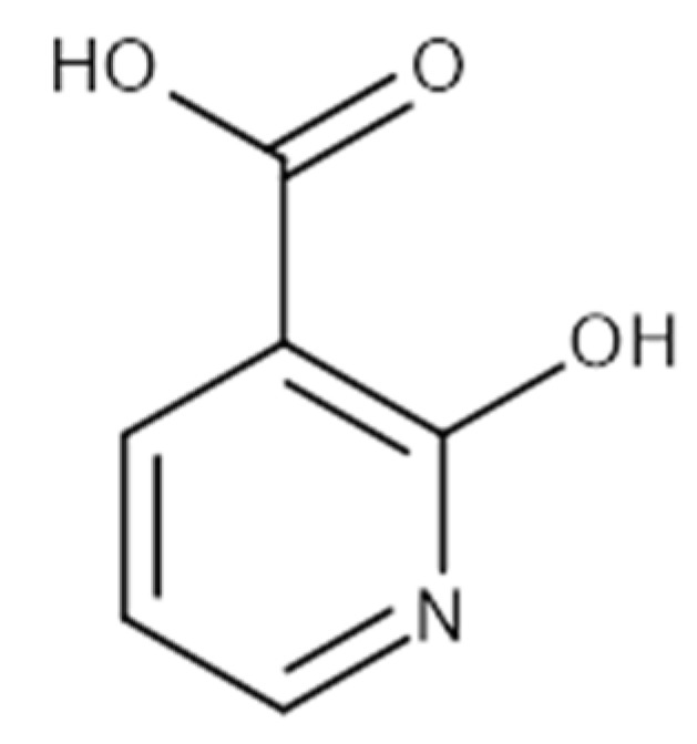
Depriving cancer cells of sufficient NAD levels, mainly through interfering with their NAD-producing capacity, has been conceived as a promising anti-cancer strategy. Numerous inhibitors of the NAD-producing enzyme, nicotinamide phosphoribosyltransferase (NAMPT), have been developed over the past two decades. However, their limited anti-cancer activity in clinical trials raised the possibility that cancer cells may also exploit alternative NAD-producing enzymes. Recent studies show the relevance of nicotinic acid phosphoribosyltransferase (NAPRT), the rate-limiting enzyme of the Preiss-Handler NAD-production pathway for a large group of human cancers. We demonstrated that the NAPRT inhibitor 2-hydroxynicotinic acid (2-HNA) cooperates with the NAMPT inhibitor FK866 in killing NAPRT-proficient cancer cells that were otherwise insensitive to FK866 alone. Despite this emerging relevance of NAPRT as a potential target in cancer therapy, very few NAPRT inhibitors exist. Starting from a high-throughput virtual screening approach, we were able to identify and annotate two additional chemical scaffolds that function as NAPRT inhibitors. These compounds show comparable anti-cancer activity to 2-HNA and improved predicted aqueous solubility, in addition to demonstrating favorable drug-like profiles.
Keywords: NAD; NAD synthesis; NAMPT; NAPRT inhibitors; anti-cancer agents; cancer metabolism; in silico drug design.
Conflict of interest statement
The authors declare no conflict of interest.
Figures


Similar articles
-
Inhibitors of NAD+ Production in Cancer Treatment: State of the Art and Perspectives.Int J Mol Sci. 2024 Feb 8;25(4):2092. doi: 10.3390/ijms25042092. Int J Mol Sci. 2024. PMID: 38396769 Free PMC article. Review.
-
Structure-Based Identification and Biological Characterization of New NAPRT Inhibitors.Pharmaceuticals (Basel). 2022 Jul 12;15(7):855. doi: 10.3390/ph15070855. Pharmaceuticals (Basel). 2022. PMID: 35890155 Free PMC article.
-
Discovery of a novel NAMPT inhibitor that selectively targets NAPRT-deficient EMT-subtype cancer cells and alleviates chemotherapy-induced peripheral neuropathy.Theranostics. 2023 Sep 11;13(14):5075-5098. doi: 10.7150/thno.85356. eCollection 2023. Theranostics. 2023. PMID: 37771778 Free PMC article.
-
Nicotinic Acid Phosphoribosyltransferase Regulates Cancer Cell Metabolism, Susceptibility to NAMPT Inhibitors, and DNA Repair.Cancer Res. 2017 Jul 15;77(14):3857-3869. doi: 10.1158/0008-5472.CAN-16-3079. Epub 2017 May 15. Cancer Res. 2017. PMID: 28507103
-
Advances in NAD-Lowering Agents for Cancer Treatment.Nutrients. 2021 May 14;13(5):1665. doi: 10.3390/nu13051665. Nutrients. 2021. PMID: 34068917 Free PMC article. Review.
Cited by
-
A Versatile Continuous Fluorometric Enzymatic Assay for Targeting Nicotinate Phosphoribosyltransferase.Molecules. 2023 Jan 18;28(3):961. doi: 10.3390/molecules28030961. Molecules. 2023. PMID: 36770640 Free PMC article.
-
Properly Substituted Benzimidazoles as a New Promising Class of Nicotinate Phosphoribosyltransferase (NAPRT) Modulators.Pharmaceuticals (Basel). 2023 Jan 27;16(2):189. doi: 10.3390/ph16020189. Pharmaceuticals (Basel). 2023. PMID: 37259338 Free PMC article.
-
Inhibitors of NAD+ Production in Cancer Treatment: State of the Art and Perspectives.Int J Mol Sci. 2024 Feb 8;25(4):2092. doi: 10.3390/ijms25042092. Int J Mol Sci. 2024. PMID: 38396769 Free PMC article. Review.
-
Targeting NAD+ metabolism: dual roles in cancer treatment.Front Immunol. 2023 Dec 5;14:1269896. doi: 10.3389/fimmu.2023.1269896. eCollection 2023. Front Immunol. 2023. PMID: 38116009 Free PMC article. Review.
-
Biological Functions and Therapeutic Potential of NAD+ Metabolism in Gynecological Cancers.Cancers (Basel). 2024 Sep 5;16(17):3085. doi: 10.3390/cancers16173085. Cancers (Basel). 2024. PMID: 39272943 Free PMC article. Review.
References
Grants and funding
- AIRC; IG#17736 and #22098) to A. Nencioni and (AIRC; IG#19172) to A. Del Rio/Italian Association for Cancer Research
- grant agreement no.813284/the European Union's Horizon 2020 research and innovation programme under the Marie Skłodowska-Curie
- (PE-2016-02362694 and PE-2016-02363073)/the BC161452P1 grant of the Breast Cancer Research Program (U.S. Department of Defense) to A. Nencioni and the Italian Ministry of Health
LinkOut - more resources
Full Text Sources
Miscellaneous
