. 2022 Jul 14;15(7):863.
doi: 10.3390/ph15070863.

The Role of Interventional Irisin on Heart Molecular Physiology

Affiliations

The Role of Interventional Irisin on Heart Molecular Physiology

Foad Alzoughool et al. Pharmaceuticals (Basel). .

Abstract

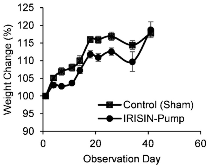
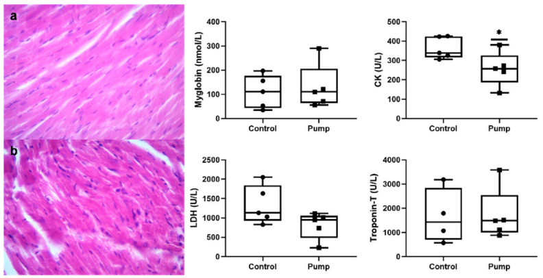
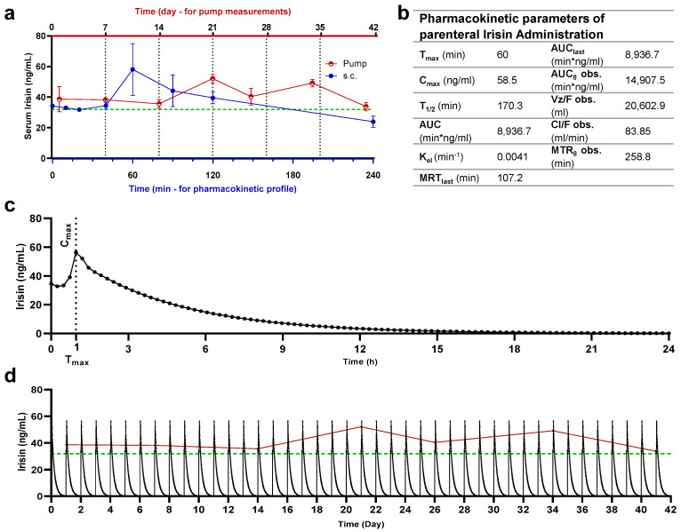
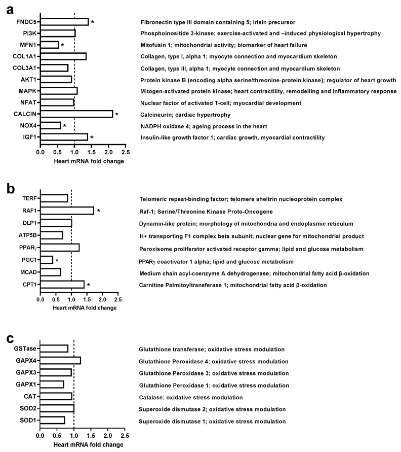
Irisin, encoded by the FNDC5 (fibronectin type III domain containing 5) gene, is a novel myokine that has been implicated as an essential mediator of exercise benefits. Effects of irisin on heart physiology is still ambiguous. This study aimed to evaluate the impact of exogenous administration of irisin on heart physiology and the pharmacokinetic profile of pump-administered irisin. To do so, Sprague Dawley rats were implanted with an irisin-loaded osmotic pump (5 μg/kg/day) for 42 days, and other animals were administered with single bolus subcutaneous injections of irisin (5 µg/kg). Body weights and blood samples were collected weekly for 42 days for serum irisin quantification and histopathology. Clinical biochemistry analyses were performed. Heart mRNA expression was assessed in 26 selected genes. Chronic interventional exogenous irisin significantly reduced body weight without affecting the heart myocyte size and significantly reduced creatine kinase enzyme level. Blood CBC, serum biochemistry, and heart morphology were normal. Gene expression of FNCD5, Raf1, CPT1, IGF-1, and CALCIN, encoding for heart physiology, increased while PGC1, Nox4, and Mfn1 significantly decreased. Nevertheless, irisin increased the expression of cardioprotective genes and inhibited some genes that harm heart physiology. Administration of irisin promotes myocardial functions and could be translated into clinical settings after preclinical profiling.

Keywords: gene expression; heart physiology; irisin; myocytes.


Conflict of interest statement

The authors declare no conflict of interest.

Figures

Figure 1
Figure 1
Percent weight change of rats administered with irisin via the osmotic pump (n = 10). Irisin reduced body weight up to 5% from day 7 and up to day 34 of administration. For example, average body weight dropped from around 209 g to 196 g at day 21 of administration.
Figure 2
Figure 2
Morphological and functional evaluation of cardiac and hind muscle of irisin-treated rats. Cardiac tissue sections of (a) control and (b) irisin (pump)-treated animals (×40). Levels of muscle activity parameters including myoglobin; CK: creatine kinase; LDH: lactate dehydrogenase, and troponin-T were analyzed in the serum of control and irisin-treated animals. * p-value < 0.05.
Figure 3
Figure 3
Relative and simulation-generated pharmacokinetic profiles of irisin. (a) Serum sampling and irisin quantification of irisin administered via the osmotic pump (upper x-axis—red) and subcutaneous route (bottom x-axis—blue) in Sprague Dawley rats, as well as baseline levels measured in control animals (dashed line—green). Irisin quantification was made at specific time intervals after administration: weekly during 42 days of osmotic pump release for rats receiving irisin via a pump and 4 h from the subcutaneous injection (min); (b) pharmacokinetic parameters of irisin in Sprague Dawley rats receiving irisin via the subcutaneous route; (c) simulated kinetic profile of single-dose subcutaneous administration; (d) repeated for 42 days with pump readings plotted with baseline levels of control animals (dashed line—green) and pump-released irisin levels (continuous line—red).
Figure 4
Figure 4
Effect of chronic administration of exogenous irisin (5 μg /kg/day) for 42 days on heart mRNA levels (n = 5). (a): Genes encoding for myocyte homeostasis and activity; (b): genes encoding for metabolism and mitochondrial activity; (c): genes encoding against oxidative stress. * p < 0.05 in comparison to the control group.

References

    1. Fagard R. Athlete’s heart. Heart. 2003;89:1455–1461. doi: 10.1136/heart.89.12.1455. - DOI - PMC - PubMed
    1. Maron B.J. Sudden Death in Young Athletes. N. Engl. J. Med. 2003;349:1064–1075. doi: 10.1056/NEJMra022783. - DOI - PubMed
    1. Morganroth J., Maron B.J., Henry W.L., Epstein S.E. Comparative Left Ventricular Dimensions in Trained Athletes. Ann. Intern. Med. 1975;82:521–524. doi: 10.7326/0003-4819-82-4-521. - DOI - PubMed
    1. Bueno O.F., De Windt L.J., Tymitz K.M., Witt S.A., Kimball T.R., Klevitsky R., Hewett T.E., Jones S., Lefer D.J., Peng C., et al. The MEK1-ERK1/2 signaling pathway promotes compensated cardiac hypertrophy in transgenic mice. EMBO J. 2000;19:6341–6350. doi: 10.1093/emboj/19.23.6341. - DOI - PMC - PubMed
    1. Cittadini A., Strömer H., Katz S.E., Clark R., Moses A.C., Morgan J.P., Douglas P.S. Differential Cardiac Effects of Growth Hormone and Insulin-like Growth Factor1 in the Rat. Circulation. 1996;93:800–809. doi: 10.1161/01.CIR.93.4.800. - DOI - PubMed