. 2022 Jul 19;15(7):893.
doi: 10.3390/ph15070893.

Pennogenin-3- O-α- L-Rhamnopyranosyl-(1→2)-[α- L-Rhamnopyranosyl-(1→3)]-β- D-Glucopyranoside (Spiroconazol A) Isolated from Dioscorea bulbifera L. var. sativa Induces Autophagic Cell Death by p38 MAPK Activation in NSCLC Cells

Affiliations

Pennogenin-3- O-α- L-Rhamnopyranosyl-(1→2)-[α- L-Rhamnopyranosyl-(1→3)]-β- D-Glucopyranoside (Spiroconazol A) Isolated from Dioscorea bulbifera L. var. sativa Induces Autophagic Cell Death by p38 MAPK Activation in NSCLC Cells

Yo Sook Ki et al. Pharmaceuticals (Basel). .

Abstract

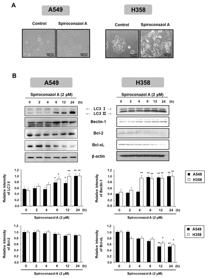
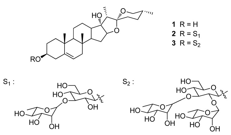
In our previous study, we reported the isolation of pennogenin-3-O-α-L-rhamnopyranosyl-(1→2)-[α-L-rhamnopyranosyl-(1→3)]-β-D-glucopyranoside (spiroconazol A), a steroidal saponin, from the flowers of Dioscorea bulbifera L. var. sativa. In the present study, we aimed to investigate the effects of spiroconazol A on autophagy and its underlying mechanisms in A549 and NCI-H358 human non-small cell lung cancer (NSCLC) cells. Spiroconazol A inhibited the proliferation of NSCLC cells in a concentration- and time-dependent manner. To determine the type of programmed cell death induced by spiroconazol A, we performed a characterization of apoptosis in spiroconazol A-treated A549 cells. Our results showed that spiroconazol A significantly suppressed A549 cell viability but did not influence cell apoptosis because phosphatidylserine and caspase activation were not detected. Furthermore, spiroconazol A treatment upregulated the expression of LC3-II and autophagy-related Beclin-1 protein, suggesting that spiroconazol A induces autophagy in A549 cells. Moreover, spiroconazol A activated the phosphorylation of p38 mitogen-activated protein kinase (MAPK) but did not affect the phosphorylation of Janus kinase or ERK1/2. Notably, SB203580, a p38 MAPK inhibitor, had a significant inhibitory effect on spiroconazol A-induced autophagic cell death in A549 cells. Our results indicated that spiroconazol A-induced autophagy is dependent on p38 MAPK signaling and has potential as a therapeutic target in NSCLC.

Keywords: autophagy; cell death; non-small cell lung cancer (NSCLC); p38 MAPK; spiroconazol A.


Conflict of interest statement

The authors declare no conflict of interest.

Figures

Figure 1
Figure 1
Non-apoptotic features were presented by spiroconazol A in A549 cells. (A) Cells were exposed to spiroconazol A (2 μM) and were stained with the PI solution. For detection of sub-G1, indicating cell death, cells were determined by flow cytometry. (B) After treatment with spiroconazol A (2 μM) for the indicated times, cells were stained with FITC-conjugated Annexin V and PI (Cis: cisplatin) and then detected by flow cytometry. (C) After treatment with 50 μM z-VAD-fmk (broad caspase inhibitor) for 1 h, cells were treated with spiroconazol A (2 μM) for 24 h. Cells were stained with the PI solution and then examined by flow cytometry. Experiments were repeated at least three times, and data are expressed as mean ± S.D. * p < 0.05, ** p < 0.01, and *** p < 0.001 vs. control group by the Student’s t-test.
Figure 2
Figure 2
Autophagy induced by spiroconazol A in NSCLC cells. (A) Representative images showed the morphological changes in spiroconazol A (2 μM)-treated NSCLC cells using an OLYMPUS IX51 inverted microscope (Southend-on-Sea, Essex, UK). (B) Protein expression of cells exposed to 2 μM of spiroconazol A, using Western blot analysis. β-actin was utilized as an internal control. The relative optical density ratio was determined using a densitometric analysis program (Bio-Rad Quantity One® Software, version 4.6.3 (Basic), Bio-Rad Laboratories Inc., CA, USA), normalized to the internal control. * p < 0.05, ** p < 0.01 vs. untreated A549 cells, by the Student’s t-test.
Figure 3
Figure 3
Cell death by spiroconazol A-induced autophagy. (A) The pEGFP-LC3-II-transfected A549 cells were treated with 2 μM of spiroconazol A for 24 h; immunofluorescence of pEGFP-LC3-II was detected by confocal fluorescence microscopy. After pretreatment with 10 mM of 3-MA for 1 h, cells were treated with 2 μM of spiroconazol A for 24 h and then examined by (B) Western blot analysis and (C) PI staining, respectively. Experiments were repeated at least three times, and data are expressed as mean ± S.D. The relative optical density ratio was determined using a densitometric analysis program (Bio-Rad Quantity One® Software, version 4.6.3 (Basic), Bio-Rad Laboratories Inc., CA, USA), normalized to the internal control. * p < 0.05, *** p < 0.001 vs. untreated A549 cells and # p < 0.05, ### p < 0.001 vs. spiroconazol A-treated A549 cells, by the Student’s t-test.
Figure 4
Figure 4
The p38 MAPK kinase signaling pathway is required in spiroconazol A-induced autophagic cell death. (A) After treatment with 2 μM of spiroconazol A for the indicated times, cells were examined by Western blot analysis. After pretreatment with 40 μM of SB203580 (p38 MAPK inhibitor), cells were treated with spiroconazol A for 24 h and then examined by (B) Western blot analysis and (C) PI staining, respectively. Experiments were repeated at least three times, and data are expressed as mean ± S.D. The relative optical density ratio was determined using a densitometric analysis program (Bio-Rad Quantity One® Software, version 4.6.3 (Basic), Bio-Rad Laboratories Inc., CA, USA), normalized to the internal control. * p < 0.05, ** p < 0.01, *** p < 0.001 vs. untreated A549 cells and # p < 0.05, ### p < 0.001 vs. spiroconazol A-treated A549 cells, by the Student’s t-test.
Figure 5
Figure 5
Chemical structures of pennogenin (1), mannioside A (2), and spiroconazol A (3) isolated from D. bulbifera L. var. sativa.

References

    1. Shen Q., Qu J., Sheng L., Gao Q., Zhou J. Case Report: Transformation From Non-Small Cell Lung Cancer to Small Cell Lung Cancer During Anti-PD-1 Therapy: A Report of Two Cases. Front. Oncol. 2021;11:619371. doi: 10.3389/fonc.2021.619371. - DOI - PMC - PubMed
    1. Tungsukruthai S., Reamtong O., Roytrakul S., Sukrong S., Vinayanwattikun C., Chanvorachote P. Targeting AKT/mTOR and Bcl-2 for Autophagic and Apoptosis Cell Death in Lung Cancer: Novel Activity of a Polyphenol Compound. Antioxidants. 2021;10:534. doi: 10.3390/antiox10040534. - DOI - PMC - PubMed
    1. Zhao R., Chen M., Jiang Z., Zhao F., Xi B., Zhang X., Fu H., Zhou K. Platycodin-D Induced Autophagy in Non-Small Cell Lung Cancer Cells via PI3K/Akt/mTOR and MAPK Signaling Pathways. J. Cancer. 2015;6:623–631. doi: 10.7150/jca.11291. - DOI - PMC - PubMed
    1. Azad M.B., Chen Y., Henson E.S., Cizeau J., McMillan-Ward E., Israels S.J., Gibson S.B. Hypoxia induces autophagic cell death in apoptosis-competent cells through a mechanism involving BNIP3. Autophagy. 2008;4:195–204. doi: 10.4161/auto.5278. - DOI - PMC - PubMed
    1. Kanzawa T., Kondo Y., Ito H., Kondo S., Germano I. Induction of autophagic cell death in malignant glioma cells by arsenic trioxide. Cancer Res. 2003;63:2103–2108. - PubMed

LinkOut - more resources