. 2022 Jun 30;14(7):1382.
doi: 10.3390/pharmaceutics14071382.

Biosynthetic Nanobubble-Mediated CRISPR/Cas9 Gene Editing of Cdh2 Inhibits Breast Cancer Metastasis

Affiliations

Biosynthetic Nanobubble-Mediated CRISPR/Cas9 Gene Editing of Cdh2 Inhibits Breast Cancer Metastasis

Ruru Gao et al. Pharmaceutics. .

Abstract

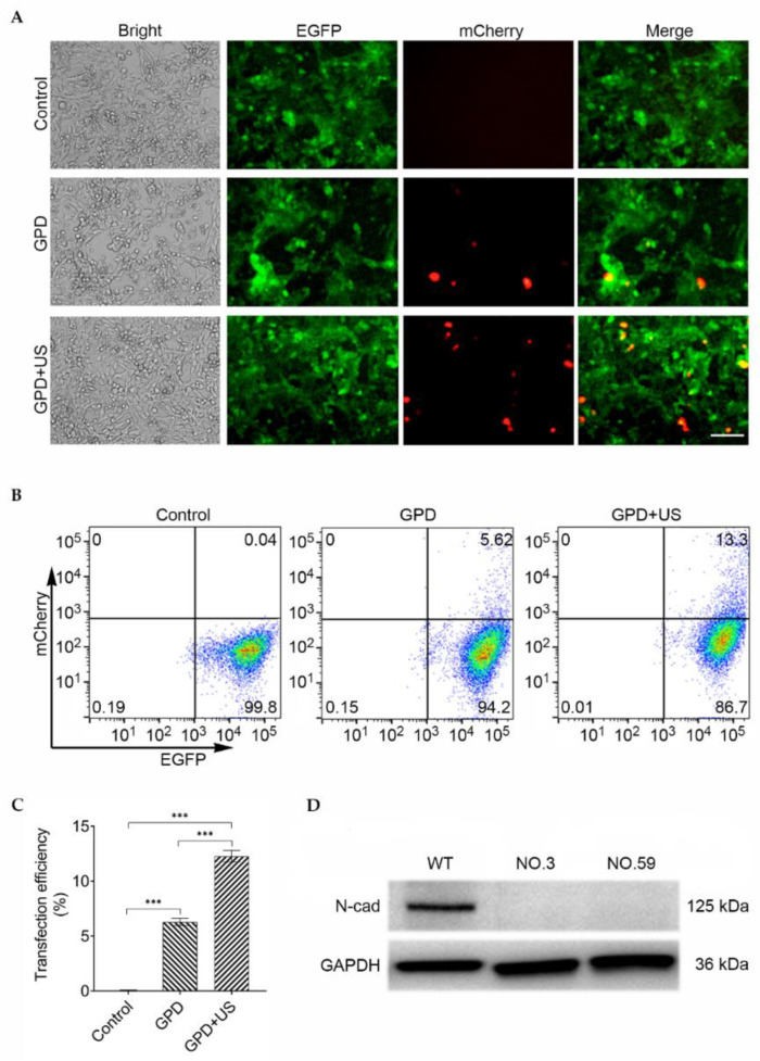
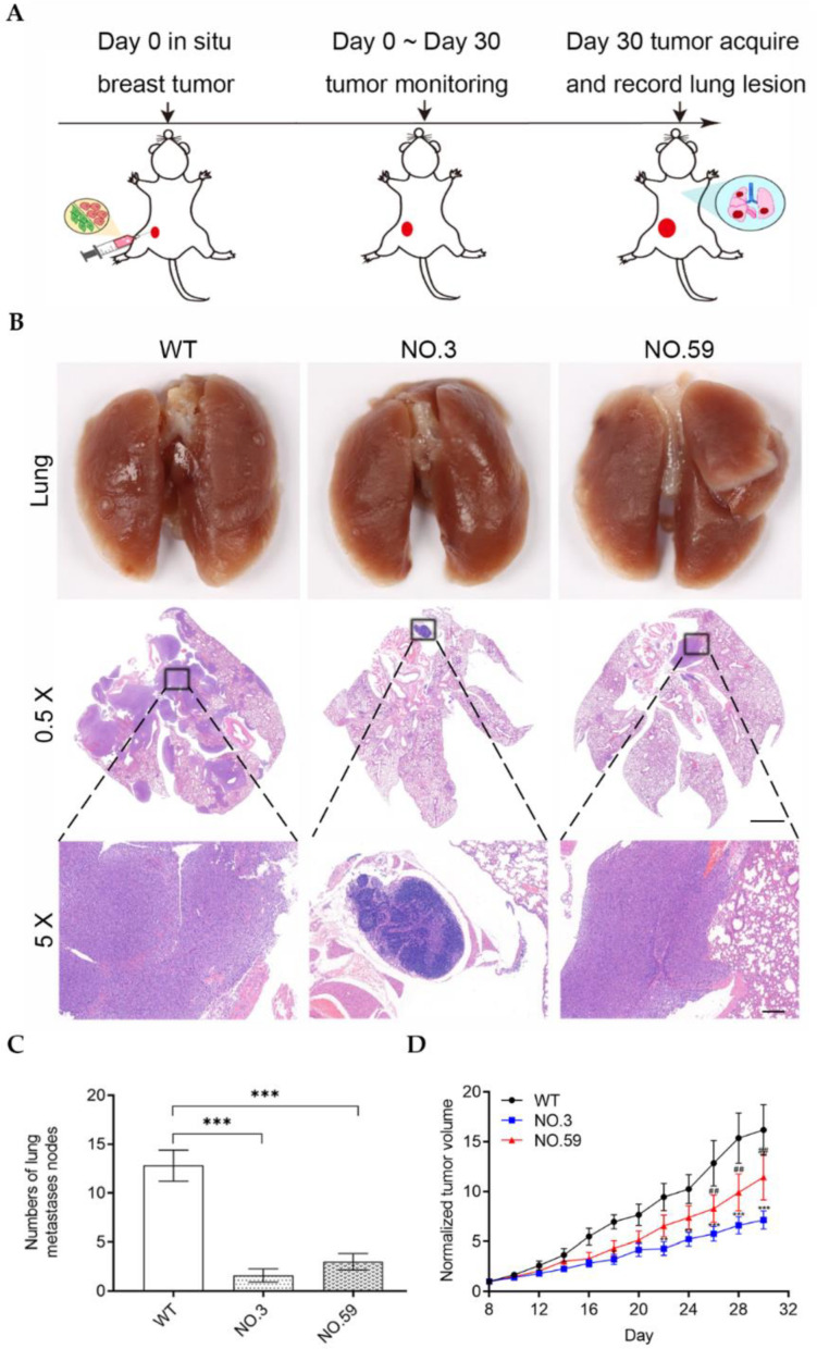
The epithelial-mesenchymal transition (EMT), a process in which epithelial cells undergo a series of biochemical changes to acquire a mesenchymal phenotype, has been linked to tumor metastasis. Here, we present a novel strategy for knocking out the EMT-related Cdh2 gene, which encodes N-cadherin through CRISPR/Cas9-mediated gene editing by an ultrasound combined with biosynthetic nanobubbles (Gas Vesicles, GVs). Polyethyleneimine were employed as a gene delivery vector to deliver sgRNA into 4T1 cells that stably express the Cas9 protein, resulting in the stable Cdh2 gene- knockout cell lines. The Western blotting assay confirmed the absence of an N-cadherin protein in these Cdh2 gene-knockout 4T1 cell lines. Significantly reduced tumor cell migration was observed in the Cdh2 gene-knockout 4T1 cells in comparison with the wild-type cells. Our study demonstrated that an ultrasound combined with GVs could effectively mediate CRISPR/Cas9 gene editing of a Cdh2 gene to inhibit tumor invasion and metastasis.

Keywords: CRISPR/Cas9; N-cadherin; epithelial–mesenchymal transition; gene editing; ultrasound.


Conflict of interest statement

The authors declare no conflict of interest. The company had no role in the design of the study; in the collection, analyses, or inter-pretation of data; in the writing of the manuscript; or in the decision to publish the results.

Figures

Figure 1
Figure 1
Schematic diagram of ultrasound-mediated CRISPR/Cas9 gene editing of Cdh2 to inhibit tumor invasion and metastasis.
Figure 2
Figure 2
(A) Fluorescence images of Cas9- and EGFP-stably expressed 4T1 cells transfected with only pU6-sgRNA (EGFP)-mCherry (control), GPD, or GPD + US (GPD: the abbreviation of GVs-PEI-DNA). Scale bar = 200 μm; (B) determination of the transfection efficiency by flow cytometry through counting of the red-emitting cells; (C) quantitative analysis of the mCherry-expressed cells from (B) (n = 3); (D) determination of the EGFP knockout efficiency (n = 3). ns denotes p > 0.05, ** p < 0.01, *** p < 0.001.
Figure 3
Figure 3
(A) Fluorescence images of Cas9- and EGFP-stably expressed 4T1 cells transfected with only pU6-sgRNA (Cdh2)-mCherry (control), GPD, or GPD + US (GPD: the abbreviation of GVs–PEI–DNA). Scale bar = 100 μm; (B) determination of the transfection efficiency by flow cytometry through the counting of the red-emitting cells in EGFP-expressed cells; (C) quantitative analysis of the mCherry-expressed cells from (B) (n = 3) *** p < 0.001. (D) Western blotting assay of the expression of N-cadherin in the wild-type (WT) and two Cdh2-KO cell lines. GAPDH was used as an internal marker.
Figure 4
Figure 4
(A) Wound healing assay of the wild-type and Cdh2-KO cell lines at 0, 12, or 24 h. Scale bar = 150 μm; (B) quantitative analysis of the recovery area of the cells from (A). (n = 3); (C) transwell cell migration assay of the wild-type and Cdh2-KO cell lines at 12, 24, or 36 h. Scale bar = 150 μm; (D) quantitative analysis of the cells migrated to the bottom chamber from (C) (n = 3). *** p < 0.001.
Figure 5
Figure 5
(A) Schematic illustration of the mouse experimental procedure for assessing the pulmonary metastasis capabilities of the Cdh2-KO cells in the orthotopic tumor model; (B) photographs of the lungs and the H&E staining of the lung sections of mice injected with the wild-type or Cdh2-KO cell lines. 0.5× magnification, Scale bar: 2 mm. 5× magnification, Scale bar: 200 μm. (C) The lung nodules were counted under an anatomical microscope. (n = 5); (D) orthotopic tumor growth curves of different groups of tumor-bearing mice. (n = 5). ‘*’: WT vs. No.3, ‘#’: WT vs. No.59. ** or ## denotes p < 0.01, *** p < 0.001.
Figure 6
Figure 6
(A) Schematic illustration of the mouse experimental procedure for assessing the pulmonary metastasis capabilities via tail intravenous injection of the Cdh2-KO cells; (B) photographs of the lungs and H&E staining of the lung sections of mice injected with the wild-type or Cdh2-KO cell lines; magnification: 0.5×, Scale bar: 2 mm. Magnification: 0.5×, Scale bar: 200 μm. (C) The lung nodules were counted under an anatomical microscope. (n = 5); (D) bodyweight change curves of the three groups of mice. (n = 5). ‘*’: WT vs. No.3, ‘#’: WT vs. No.59. ## denotes p < 0.01, *** or ### p < 0.001.

References

    1. Sun D., Cao M., Li H., He S., Chen W. Cancer burden and trends in China: A review and comparison with Japan and South Korea. Chin. J. Cancer Res. 2020;32:129–139. doi: 10.21147/j.issn.1000-9604.2020.02.01. - DOI - PMC - PubMed
    1. Siegel R.L., Miller K.D., Fuchs H.E., Jemal A. Cancer Statistics, 2021. CA Cancer J. Clin. 2021;71:7–33. doi: 10.3322/caac.21654. - DOI - PubMed
    1. Walker A., Frei R., Lawson K.R. The cytoplasmic domain of N-cadherin modulates MMP9 induction in oral squamous carcinoma cells. Int. J. Oncol. 2014;45:1699–1706. doi: 10.3892/ijo.2014.2549. - DOI - PMC - PubMed
    1. Wang Y., Zhang T., Kwiatkowski N., Abraham B.J., Lee T.I., Xie S., Yuzugullu H., Von T., Li H., Lin Z., et al. CDK7-dependent transcriptional addiction in triple-negative breast cancer. Cell. 2015;163:174–186. doi: 10.1016/j.cell.2015.08.063. - DOI - PMC - PubMed
    1. Hennessy B.T., Gonzalez-Angulo A.M., Stemke-Hale K., Gilcrease M.Z., Krishnamurthy S., Lee J.S., Fridlyand J., Sahin A., Agarwal R., Joy C., et al. Characterization of a naturally occurring breast cancer subset enriched in epithelial-to-mesenchymal transition and stem cell characteristics. Cancer Res. 2009;69:4116–4124. doi: 10.1158/0008-5472.CAN-08-3441. - DOI - PMC - PubMed