. 2022 Jul 5;14(7):1412.
doi: 10.3390/pharmaceutics14071412.

Reduction of αSYN Pathology in a Mouse Model of PD Using a Brain-Penetrating Bispecific Antibody

Affiliations

Reduction of αSYN Pathology in a Mouse Model of PD Using a Brain-Penetrating Bispecific Antibody

Sahar Roshanbin et al. Pharmaceutics. .

Abstract

Immunotherapy targeting aggregated alpha-synuclein (αSYN) is a promising approach for the treatment of Parkinson's disease. However, brain penetration of antibodies is hampered by their large size. Here, RmAbSynO2-scFv8D3, a modified bispecific antibody that targets aggregated αSYN and binds to the transferrin receptor for facilitated brain uptake, was investigated to treat αSYN pathology in transgenic mice. Ex vivo analyses of the blood and brain distribution of RmAbSynO2-scFv8D3 and the unmodified variant RmAbSynO2, as well as in vivo analyses with microdialysis and PET, confirmed fast and efficient brain uptake of the bispecific format. In addition, intravenous administration was shown to be superior to intraperitoneal injections in terms of brain uptake and distribution. Next, aged female αSYN transgenic mice (L61) were administered either RmAbSynO2-scFv8D3, RmAbSynO2, or PBS intravenously three times over five days. Levels of TBS-T soluble aggregated αSYN in the brain following treatment with RmAbSynO2-scFv8D3 were decreased in the cortex and midbrain compared to RmAbSynO2 or PBS controls. Taken together, our results indicate that facilitated brain uptake of αSYN antibodies can improve treatment of αSYN pathology.

Keywords: Parkinson’s disease (PD); alpha-synuclein (αSYN); bispecific antibody; blood-brain barrier (BBB); immunotherapy; monoclonal antibody; receptor-mediated transcytosis (RMT); transferrin receptor (TfR).


Conflict of interest statement

The authors declare no conflict of interest.

Figures

Figure 1
Figure 1
(A) Design of the bispecific RmAbSynO2-scFv8D3 with a single chain variable fragment of the mTfR binding antibody 8D3 fused to the C-terminus of the light chain with a short linker and the unmodified RmAbSynO2. (B) Blood exposure of [125I]RmAbSynO2-scFv8D3 and (C) [125I]RmAbSynO2 following intravenous (i.v.) and intraperitoneal (i.p.) injections. Blood exposure of both antibody formats was higher after i.v. compared to i.p. administration, with [125I]RmAbSynO2-scFv8D3 displaying lower total exposure. (D) Total brain concentrations of [125I]RmAbSynO2-scFv8D3 and (E) [125I]RmAbSynO2 were both higher after i.v. injections in comparison with i.p., with [125I]RmAbSynO2-scFv8D3 concentrations peaking 2–4 h post-injection and substantially higher than [125I]RmAbSynO2 at all points (NB different y-axis scale). (F) Radioactive signal in 20 µm sagittal brain cryosections from WT mice in (D,E), demonstrating that mice i.v. injected with [125I]RmAbSynO2-scFv8D3 showed the highest signals at all time points. (G) Levels of [125I]RmAbSynO2-scFv8D3 were higher in interstitial fluid (ISF) dialysate measured by microdialysis in the striatum after i.v. injections compared to after i.p. injections (mean ± SEM, p = 0.0313, n = 4/administration route). All blood and brain concentrations are expressed as % injected dose per gram tissue, normalized to bodyweight (%ID/g tissue/BW).
Figure 2
Figure 2
(A) Sagittal PET images of WT mice injected with [18F]RmAbSynO2-scFv8D3 (upper; n = 1) or [18F]RmAbSynO2 (lower; n = 1) at 0–5 min and 90–120 min post-injection. (B) Quantification of brain (whole brain except cerebellum) activity concentration (SUV) from 0–120 min after antibody injection in same animals as (A).
Figure 3
Figure 3
(A) Study design of a short-term treatment study in female L61 αSYN mice between 14-16 months of age. Mice were treated with i.v. injections 10 mg/kg RmAbSynO2-scFv8D3 or RmAbSynO2 or PBS (n = 8 per treatment group) on day 1, 3 and 4, with study termination on day 5. (B) Lower blood exposure of [125I]RmAbSynO2-scFv8D3 compared with [125I]RmAbSynO2 at 1 h, 4 h and 24 h after each injection. (C) Higher concentration of [125I]RmAbSynO2-scFv8D3 in the brain compared to [125I]RmAbSynO2 (7.1 ± 0.43 and 3.2 ± 0.16%ID/g brain/bw, respectively) upon study termination. (D) Higher brain-to-blood ratio of [125I]RmAbSynO2-scFv8D3 (0.043 ± 0.0021) in comparison with [125I]RmAbSynO2 (0.0048 ± 0.00013). All values are presented as means ± SD and analyzed with an unpaired t-test. **** p < 0.0001.
Figure 4
Figure 4
(A) Levels of total αSYN in TBS and TBS-T brain extracts measured with MJFR1/Syn-1 enzyme-linked immunosorbent assay (ELISA) measuring all forms of αSYN. No differences in total αSYN in the cortex (Cx), midbrain (Mb), and cerebellum (Cer). Reduced total αSYN in Cx in the TBS-T fraction in mice treated with RmAbSynO2-scFv8D3 in comparison with the PBS group. (B) Oligomeric αSYN levels in TBS and TBS-T extracts measured with a homogenous MJFR-14-6-4-2 ELISA. Oligomeric αSYN in the TBS fraction was increased in Cx in the RmAbSynO2-scFv8D3 group in comparison with both the RmAbSynO2- and the PBS control groups and in Mb compared to the PBS group. Oligomeric αSYN levels in the TBS-T fraction were instead decreased in Cx and Mb in the RmAbSynO2-scFv8D3 group compared to PBS controls, while Cer αSYN levels were unchanged. (C) Representative images of 20 µm cryosections stained for Iba1 in Cx, hippocampus (HC), and Cer at 60× magnification, with 4× magnifications embedded with squares representing the magnified area. No differences in immunoreactivity were noted between the treatment groups. All values are presented as means ± SD, with a one-way analysis of variance followed by Sidak’s multiple comparison test. * p < 0.05, ** p < 0.005. Scale bars: 200 µm in embedded images, 10 µm in magnified images.
Figure 5
Figure 5
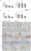
(A) ELISA quantification of sTREM2 levels in TBS and TBS-T extracts from the cortex (Cx), midbrain (Mb), and cerebellum (Cer), following treatment with RmAbSynO2-scFv8D3 or RmAbSynO2 in comparison with PBS. Both antibody treatment groups displayed increased sTREM2 levels in both TBS and TBS-T extract of most examined regions. (B) Representative images of 20 µm cryosections stained for Iba1 in Cx, hippocampus (HC), and Cer at 60× magnification, with 4× magnifications embedded with squares representing the magnified area, showing increased immunoreactivity on sections from mice treated with both antibody formats. All values are presented as means ± SD, with a one-way analysis of variance followed by Sidak’s multiple comparison test. * p < 0.05, ** p < 0.005, *** p < 0.001. Scale bars: 200 µm in embedded images, 10 µm in magnified images.

Similar articles

Cited by

References

    1. Polymeropoulos M.H., Lavedan C., Leroy E., Ide S.E., Dehejia A., Dutra A., Pike B., Root H., Rubenstein J., Boyer R., et al. Mutation in the alpha-synuclein gene identified in families with Parkinson’s disease. Science. 1997;276:2045–2047. doi: 10.1126/science.276.5321.2045. - DOI - PubMed
    1. Krüger R., Kuhn W., Müller T., Woitalla D., Graeber M., Kösel S., Przuntek H., Epplen J.T., Schöls L., Riess O. Ala30Pro mutation in the gene encoding alpha-synuclein in Parkinson’s disease. Nat. Genet. 1998;18:106–108. doi: 10.1038/ng0298-106. - DOI - PubMed
    1. Spillantini M.G., Schmidt M.L., Lee V.M., Trojanowski J.Q., Jakes R., Goedert M. Alpha-synuclein in Lewy bodies. Nature. 1997;388:839–840. doi: 10.1038/42166. - DOI - PubMed
    1. Bohnen N.I., Albin R.L. The cholinergic system and Parkinson disease. Behav. Brain Res. 2011;221:564–573. doi: 10.1016/j.bbr.2009.12.048. - DOI - PMC - PubMed
    1. Schapira A.H., Emre M., Jenner P., Poewe W. Levodopa in the treatment of Parkinson’s disease. Eur. J. Neurol. 2009;16:982–989. doi: 10.1111/j.1468-1331.2009.02697.x. - DOI - PubMed

LinkOut - more resources