Review
. 2022 Jul 13;10(7):1120.
doi: 10.3390/vaccines10071120.

Advances in Infectious Disease Vaccine Adjuvants

Affiliations
Review

Advances in Infectious Disease Vaccine Adjuvants

Jingyi Fan et al. Vaccines (Basel). .

Abstract

Vaccines are one of the most significant medical interventions in the fight against infectious diseases. Since their discovery by Edward Jenner in 1796, vaccines have reduced the worldwide transmission to eradication levels of infectious diseases, including smallpox, diphtheria, hepatitis, malaria, and influenza. However, the complexity of developing safe and effective vaccines remains a barrier for combating many more infectious diseases. Immune stimulants (or adjuvants) are an indispensable factor in vaccine development, especially for inactivated and subunit-based vaccines due to their decreased immunogenicity compared to whole pathogen vaccines. Adjuvants are widely diverse in structure; however, their overall function in vaccine constructs is the same: to enhance and/or prolong an immunological response. The potential for adverse effects as a result of adjuvant use, though, must be acknowledged and carefully managed. Understanding the specific mechanisms of adjuvant efficacy and safety is a key prerequisite for adjuvant use in vaccination. Therefore, rigorous pre-clinical and clinical research into adjuvant development is essential. Overall, the incorporation of adjuvants allows for greater opportunities in advancing vaccine development and the importance of immune stimulants drives the emergence of novel and more effective adjuvants. This article highlights recent advances in vaccine adjuvant development and provides detailed data from pre-clinical and clinical studies specific to infectious diseases. Future perspectives into vaccine adjuvant development are also highlighted.

Keywords: immunological adjuvants; infectious diseases; pre-clinical and clinical trials.


Conflict of interest statement

The authors declare no conflict of interest.

Figures

Figure 1
Figure 1
Schematic of an immune response following vaccination. In adaptive immunity, antigens combined with adjuvants are delivered to and bind with naïve dendritic cells, forming antigen-presenting cells (mature dendritic cells), which are recognized by major histocompatibility complex (MHC) class I and MHC-II, thereby binding with T-cell receptors on naïve CD8+ cells and naïve CD4+ T cells, respectively. Naïve CD4+ cells stimulate the production of Th1 (or Th2) responsible for the secretion of different cytokines, and induction of cellular and humoral immunity, respectively.
Figure 2
Figure 2
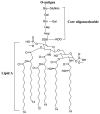
Structure of E. coli lipopolysaccharide. Abbreviations Gal: D-galactose; Glu: D-glucose; Hep: L-glycero-D-manno-heptose; KDO: 3-deoxy-D-manno-oct-2-ulosonic acid; P: phosphate.
Figure 3
Figure 3
Chemical components of MF59®, where MF59® is composed of squalene and two surfactants (Span 85 and Tween 80). MF59® and AS03 not only have similar components, but also analogous composition. These are mixed in an oil phase giving oil-in-water emulsions.
Figure 4
Figure 4
Schematic of a virus-like particle (VLP), where the surface is made up of antigens (e.g., capsid proteins) that have been embedded into the natural lipid bilayer (surrounding cell membrane) to form enveloped VLPs. (a) Non-enveloped VLPs contain single or multiple capsid proteins only. Neither type of VLP contains infectious material (e.g., DNA/RNA). (b) The formulation of double layer enveloped VLPs is related to the multiple glycoproteins on their surface.
Figure 5
Figure 5
Schematic virosome adjuvant. Virosomes are synthesised from a phospholipid bilayer (similar to that of a liposome) where approved influenza vaccines are used the virosomes virus structure allowing for conjugation with influenza surface protein hemagglutinin (HA) and neuraminidase (NA).
Figure 6
Figure 6
Liposome schematic.
Figure 7
Figure 7
Schematic polymeric nanoparticles.
Figure 8
Figure 8
Structure of α-Galactosylceramide.
Figure 9
Figure 9
QS-21 adjuvant structure.
Figure 10
Figure 10
Schematic of immune-stimulating complexes (ISCOMs).
Figure 11
Figure 11
(a) Components of adjuvant system AS03 (assembled from squalene, polysorbate 80 and α-tocopherola [a type of vitamin E]). (b) MPL structure of adjuvant system AS04 as a TLR4 agonist which is mixed with alum.

References

    1. Baker R.E., Mahmud A.S., Miller I.F., Rajeev M., Rasambainarivo F., Rice B.L., Takahashi S., Tatem A.J., Wagner C.E., Wang L.F., et al. Infectious disease in an era of global change. Nat. Rev. Microbiol. 2022;20:193–205. doi: 10.1038/s41579-021-00639-z. - DOI - PMC - PubMed
    1. Talisuna A.O., Bloland P., D’Alessandro U. History, dynamics, and public health importance of malaria parasite resistance. Clin. Microbiol. Rev. 2004;17:235–254. doi: 10.1128/CMR.17.1.235-254.2004. - DOI - PMC - PubMed
    1. Andreano E., D’Oro U., Rappuoli R., Finco O. Vaccine Evolution and Its Application to Fight Modern Threats. Front. Immunol. 2019;10:1722. doi: 10.3389/fimmu.2019.01722. - DOI - PMC - PubMed
    1. Dworetzky M., Cohen S., Mullin D. Prometheus in Gloucestershire: Edward Jenner, 1749-1823. J. Allergy Clin. Immunol. 2003;112:810–814. doi: 10.1016/S0091-6749(03)02017-7. - DOI - PubMed
    1. Azuar A., Jin W., Mukaida S., Hussein W.M., Toth I., Skwarczynski M. Recent advances in the development of peptide vaccines and their delivery systems against Group A Streptococcus. Vaccines. 2019;7:58. doi: 10.3390/vaccines7030058. - DOI - PMC - PubMed

LinkOut - more resources