. 2022 Jul 2;14(7):1465.
doi: 10.3390/v14071465.

Pseudorabies Virus Tegument Protein UL13 Suppresses RLR-Mediated Antiviral Innate Immunity through Regulating Receptor Transcription

Affiliations

Pseudorabies Virus Tegument Protein UL13 Suppresses RLR-Mediated Antiviral Innate Immunity through Regulating Receptor Transcription

Ningning Zhao et al. Viruses. .

Abstract

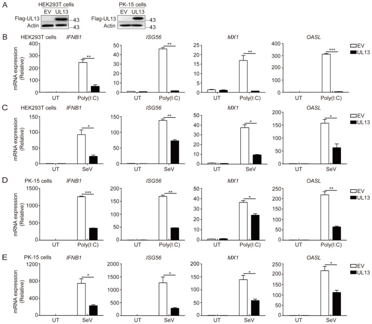
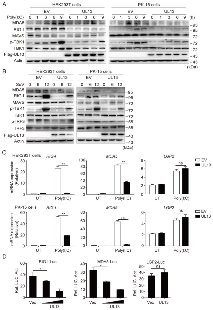
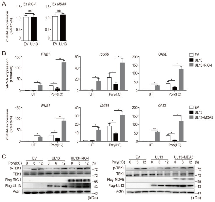
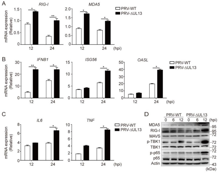
Pseudorabies virus (PRV) has evolved various strategies to escape host antiviral immune responses. However, it remains unclear whether and how PRV-encoded proteins modulate the RIG-I-like receptor (RLR)-mediated signals for immune evasion. Here, we show that the PRV tegument protein UL13 functions as an antagonist of RLR-mediated antiviral responses via suppression of the transcription of RIG-I and MDA5, but not LGP2. UL13 overexpression significantly inhibits both the mRNA and protein levels of RIG-I and MDA5, along with RIG-I- or MDA5-mediated antiviral immune responses, whereas overexpression of RIG-I or MDA5 counteracts such UL13-induced suppression. Mechanistically, UL13 suppresses the expression of RIG-I and MDA5 by inhibiting activation of the transcription factor NF-κB. Consequently, overexpression of p65 promotes the activation of RIG-I and MDA5 promoters. Moreover, deletion of the p65-binding sites in the promoters of RIG-I or MDA5 abolishes the suppression role of UL13. As a result, mutant PRV lacking UL13 elicits stronger host antiviral immune responses than PRV-WT. Hence, our results provide a novel functional role of UL13-induced suppression of host antiviral immunity through modulating receptors' transcription.

Keywords: MDA5; NF-κB; RIG-I; pseudorabies virus (PRV); tegument protein UL13.


Conflict of interest statement

The authors declare no conflict of interest.

Figures

Figure 1
Figure 1
PRV UL13 negatively regulated RLR-mediated expression of type-I IFN and ISGs. (A) The expression of Flag-tagged UL13 in HEK293T or PK-15 cell lines was verified by immunoblotting; β-actin served as a loading control. (BE) Quantitative real-time PCR (qPCR) analysis of IFNB1 and downstream ISG (ISG56, MX1, and OASL) mRNA expression in UL13-HEK293T cells (B,C) or UL13-PK-15 cells (D,E), and control cells left untreated (UT) or transfected with 1 µg/mL of poly(I:C) (B,D) for 6 h or infected with SeV (MOI = 1) (C,E) for 12 h. Data are pooled from three independent experiments (mean ± SEM); *, p < 0.05; **, p < 0.01; ***, p < 0.001 (Student’s t-test).
Figure 2
Figure 2
UL13 inhibits transcription of RIG-I and MDA5 to suppress RLR-mediated antiviral responses. (A,B) Immunoblotting analysis of RIG-I, MDA5, MAVS, phosphorylated (Ser172) and total TBK1, and phosphorylated (Ser396) and total IRF3 in whole-cell lysates of UL13-HEK293T or UL13-PK-15 cells and control cells stimulated with poly(I:C) (A) or infected with SeV (MOI = 1, B) for the indicated times (above lanes); β-actin served as a loading control. (C) qPCR analysis of the mRNA levels of RIG-I, MDA5, and LGP2 in UL13-HEK293T cells, UL13-PK-15 cells, or control cells left untreated (UT) or transfected with poly(I:C) for 6 h. (D) Luciferase activities in HEK293T cells co-transfected with RIG-I, MDA5, or LGP2 promoter-driven luciferase reporters (50 ng) and plasmids encoding UL13 (concentration 150 ng, 300 ng) or empty vectors. Twenty-four hours after transfection, cell lysates were analyzed for luciferase activity. Data are representative of three independent experiments (A,B), or are pooled from three independent experiments (C,D, mean ± SD); *, p < 0.05; **, p < 0.01; ***, p < 0.001; ns, not significant (Student’s t-test).
Figure 3
Figure 3
Enforced expression of RIG-I or MDA5 rescues UL13-suppressed antiviral immune responses. (A) qPCR analysis of RIG-I and MDA5 mRNA expression in UL13-HEK293T cells or control cells transfected with plasmids encoding Flag-RIG-I or Flag-MDA5 for 24 h. (B) qPCR analysis of IFNB1 and downstream ISG (ISG56 and OASL) mRNA expression in cells transfected with plasmids encoding Flag-RIG-I, Flag-MDA5, or an empty vector for 24 h following poly(I:C) transfection for 6 h. (C) Immunoblotting analysis of phosphorylated (Ser172) and total TBK1, Flag-RIG-I or Flag-MDA5, and Flag-UL13 in UL13-HEK293T cells transfected with plasmids encoding Flag-RIG-I, Flag-MDA5, or an empty vector for 24 h following poly(I:C) transfection for the additional indicated periods. Data are pooled from three independent experiments (A,B, mean ± SD) or representative of three independent experiments (C); *, p < 0.05; **, p < 0.01; ns, not significant (Student’s t-test).
Figure 4
Figure 4
UL13 represses transcription of RIG-I and MDA5 by regulating NF-κB activation. (A) qPCR analysis of mRNA expression of IFNB1 and downstream ISG (ISG56 and MX1) in UL13-HEK293T cells or control cells. Cells were left untreated or transfected with poly(I:C) for 6 h, or pretreated with NF-κB inhibitor (BAY11-7082, 10 µM), JNK inhibitor (SP600125, 10 µM), MEK/ERK inhibitor (U0126, 10 µM), or p38 inhibitor (SB203580, 10 µM) for 1 h, followed by poly(I:C) stimulation for 6 h. (B) Immunoblotting analysis of RIG-I, phosphorylated (Ser356) and total p65, IκBα, and Flag-UL13 in whole-cell lysates of UL13-HEK293T cells or control cells transfected with or without poly(I:C) for the indicated times. (C) Immunoblotting analysis of p65 protein levels in the nucleus and cytoplasm in UL13-HEK293T cells and control cells transfected with poly(I:C) for 3 h; β-actin and Lamin B1 served as the loading controls for the cytoplasm and nucleus, respectively. (D) Immunofluorescence analysis of the nuclear translocation of p65 in UL13-HEK293T cells or control cells left untreated or transfected with poly(I:C) for 3 h. Scale bars = 20 μm. (E) Quantification of nuclear localization of p65. (F) qPCR analysis of mRNA expression of IL6 and TNF in cells as in (D). (G) Luciferase activities in UL13-HEK293T cells or control cells co-transfected with RIG-I-, MDA5-, or LGP2-promoter-driven luciferase reporters and plasmids encoding p65 or empty vectors. Twenty-four hours after transfection, cell lysates were analyzed for luciferase activity. (H) Annotation of putative p65-binding sites in RIG-I promoter or MDA5 promoter and their mutants. (I) Luciferase activities in UL13-HEK293T cells or control cells co-transfected with RIG-I- or MDA5-promoter-driven luciferase reporters or mutants with plasmid encoding p65 or empty vectors. Twenty-four hours after transfection, cell lysates were analyzed for luciferase activity. Data are pooled from three independent experiments (A,EG,I, mean ± SD) or representative of three independent experiments (B,D); *, p < 0.05; **, p < 0.01; ns, not significant (Student’s t-test).
Figure 5
Figure 5
UL13 deficiency increases expression of RLRs and promotes RLR-mediated antiviral responses. (A) qPCR analysis of RIG-I and MDA5 mRNA levels in HEK293T cells infected with PRV-WT or PRV-∆UL13 (MOI = 1) for the indicated times. (B,C) qPCR analysis the mRNA levels of IFNB1, downstream ISGs (ISG56 and OASL) (B), and expression of IL6 and TNF (C) in HEK293T cells infected with PRV-WT or PRV-∆UL13 (MOI = 1) for the indicated periods. (D) Immunoblotting analysis of MDA5, RIG-I, MAVS, phosphorylated and total TBK1, and phosphorylated and total p65 in HEK293T cells infected with PRV-WT or PRV-∆UL13 (MOI = 1). Data are pooled from three independent experiments (A, B, C, mean ± SD) or representative of three independent experiments (D); *, p < 0.05; **, p < 0.01; ns, not significant (Student’s t-test).

Similar articles

Cited by

References

    1. Laval K., Enquist L.W. The Neuropathic Itch Caused by Pseudorabies Virus. Pathogens. 2020;9:254. doi: 10.3390/pathogens9040254. - DOI - PMC - PubMed
    1. Li H., Liang R., Pang Y., Shi L., Cui S., Lin W. Evidence for interspecies transmission route of pseudorabies virus via virally contaminated fomites. Vet. Microbiol. 2020;251:108912. doi: 10.1016/j.vetmic.2020.108912. - DOI - PubMed
    1. Liu J., Chen C., Li X. Novel Chinese pseudorabies virus variants undergo extensive recombination and rapid interspecies transmission. Transbound. Emerg. Dis. 2020;67:2274–2276. doi: 10.1111/tbed.13784. - DOI - PubMed
    1. Liu Q., Wang X., Xie C., Ding S., Yang H., Guo S., Li J., Qin L., Ban F., Wang D., et al. A novel human acute encephalitis caused by pseudorabies virus variant strain. Clin. Infect. Dis. 2020;73:e3690–e3700. doi: 10.1093/cid/ciaa987. - DOI - PubMed
    1. Ai J.-W., Weng S.-S., Cheng Q., Cui P., Li Y.-J., Wu H.-L., Zhu Y.-M., Xu B., Zhang W.-H. Human Endophthalmitis Caused By Pseudorabies Virus Infection, China, 2017. Emerg. Infect. Dis. 2018;24:1087–1090. doi: 10.3201/eid2406.171612. - DOI - PMC - PubMed

Publication types

MeSH terms

LinkOut - more resources