. 2022 Jul 8;14(7):1501.
doi: 10.3390/v14071501.

Reporter Flaviviruses as Tools to Demonstrate Homologous and Heterologous Superinfection Exclusion

Affiliations

Reporter Flaviviruses as Tools to Demonstrate Homologous and Heterologous Superinfection Exclusion

Francisco J Torres et al. Viruses. .

Abstract

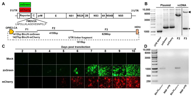
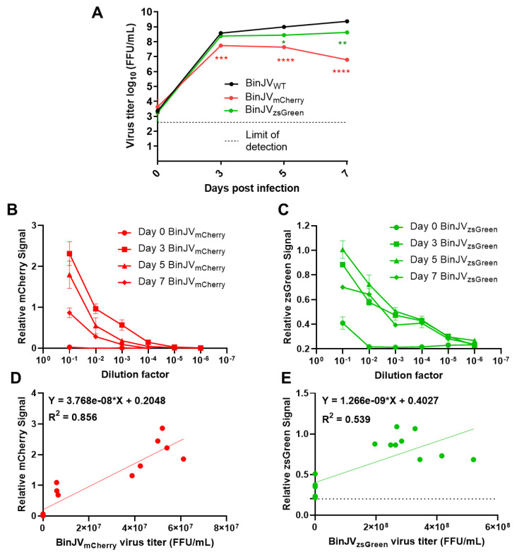
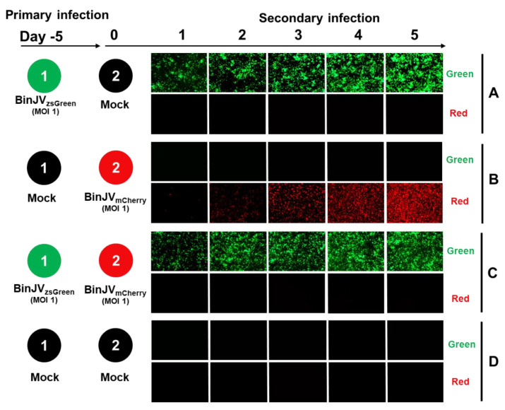
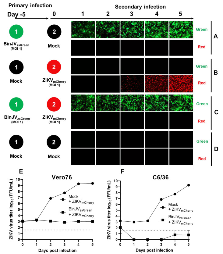
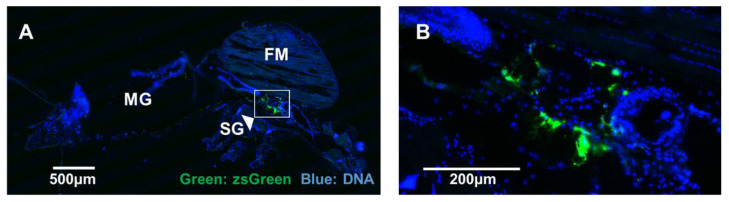
Binjari virus (BinJV) is a lineage II or dual-host affiliated insect-specific flavivirus previously demonstrated as replication-deficient in vertebrate cells. Previous studies have shown that BinJV is tolerant to exchanging its structural proteins (prM-E) with pathogenic flaviviruses, making it a safe backbone for flavivirus vaccines. Here, we report generation by circular polymerase extension reaction of BinJV expressing zsGreen or mCherry fluorescent protein. Recovered BinJV reporter viruses grew to high titres (107-8 FFU/mL) in Aedes albopictus C6/36 cells assayed using immunoplaque assays (iPA). We also demonstrate that BinJV reporters could be semi-quantified live in vitro using a fluorescence microplate reader with an observed linear correlation between quantified fluorescence of BinJV reporter virus-infected C6/36 cells and iPA-quantitated virus titres. The utility of the BinJV reporter viruses was then examined in homologous and heterologous superinfection exclusion assays. We demonstrate that primary infection of C6/36 cells with BinJVzsGreen completely inhibits a secondary infection with homologous BinJVmCherry or heterologous ZIKVmCherry using fluorescence microscopy and virus quantitation by iPA. Finally, BinJVzsGreen infections were examined in vivo by microinjection of Aedes aegypti with BinJVzsGreen. At seven days post-infection, a strong fluorescence in the vicinity of salivary glands was detected in frozen sections. This is the first report on the construction of reporter viruses for lineage II insect-specific flaviviruses and establishes a tractable system for exploring flavivirus superinfection exclusion in vitro and in vivo.

Keywords: Binjari virus; CPER; flavivirus; insect-specific viruses; reporter viruses; superinfection exclusion.


Conflict of interest statement

N.D.N., L.J.V., J.H.-P., R.A.H. and A.A.K. are inventors on a patent (WO/2018/176075) covering the Binjari chimera technology. No other authors declare competing interests.

Figures

Figure 1
Figure 1
Design and recovery of BinJV reporter viruses. (A) Schematics of BinJV and overlapping fragments and linker fragments used for CPER assembly. (B) Agarose gel electrophoresis of BinJV fragments amplified from viral cDNA, BinJVzsGreen/mCherry fragments and the UTR linker fragment from plasmids. Arrowhead in F3 lane shows correct size band, while asterisk in F3 lane shows non-specific amplification. The image for F3 is from the same gel with unrelated lanes spliced out for clarity. (C) Images of zsGreen and mCherry fluorescence of C6/36 cells transfected with reporter virus CPER were taken at 40× magnification daily from two to ten days post-transfection. (D) RT-PCR for the reporter insert of P1 BinJV recombinants propagated in C6/36 cells. RNA-only samples refer to the PCR amplification of RNA samples without the reverse transcription step. Amplicons were generated using RGene_F and RGene_R primers binding to the BinJV sequences flanking the reporter genes.
Figure 2
Figure 2
Binjari virus reporters grow to high titres in insect cells and are semi-quantifiable using fluorescent microplate readers. (A) Growth kinetics of P1 CPER generated BinJV viruses in C6/36 cells infected with WT or reporter BinJV. Cells were infected at MOI = 0.1, culture supernatant were sampled at the indicated time point, and titers were determined using iPA on C6/36 cells. Values are the means from three biological replicates ±SEM. Statistical analysis was a two-way ANOVA with Dunnett’s correction. All comparisons were to WT; * p < 0.05, ** p < 0.01, *** p < 0.001, **** p < 0.0001. Relative fluorescence units for serially diluted C6/36 culture supernatant taken from the growth kinetics and reinfected on C6/36 cells showing (B) BinJVmCherry signal and (C) BinJVzsGreen signal. The linear relationship between matched samples of fluorescence units taken from the 10−1 dilution and FFU/mL assay of (D) BinJVmCherry (E) BinJVzsGreen viruses. For the simple linear regression analysis, 15 matched samples were used. The baseline signal for zsGreen is given as a dotted line. The linear regression function and the coefficient of determination (R2) are given on both graphs.
Figure 3
Figure 3
Superinfection exclusion between BinJVzsGreen and BinJVmCherry viruses. Schematic showing the format of the infection trials and representative images of green and red fluorescence channels of C6/36 cells with (A) primary infections of BinJVzsGreen at −5 days and mock (media only) secondary infection. (B) Mock primary infection at −5 days and BinJVmCherry secondary infection. (C) Primary infections of BinJVzsGreen at −5 days, BinJVmCherry secondary infection, and (D) mock (uninfected) primary and secondary timepoints.
Figure 4
Figure 4
Superinfection exclusion of ZIKV by the insect-specific BinJV. Schematic showing the format of the infection trials and representative images of green and red fluorescence channels of C6/36 cells with (A) primary infections of BinJVzsGreen at −5 days and mock (media only) secondary infection. (B) Mock primary infection at −5 days and ZIKVmCherry secondary infection. (C) Primary infections of BinJVzsGreen at −5 days, ZIKVmCherry secondary infection, and (D) mock (uninfected) primary and secondary timepoints. iPA assay of culture supernatant from the indicated time points of experiments (B,C) titred on Vero76 (E) and Ae. albopictus (F) cells. Values are the means from three replicates ±SEM. The dotted line is the limit of detection.
Figure 5
Figure 5
Microinjection of Aedes aegypti with BinJVzsGreen reporter virus. (A) zsGreen signal (green) in a representative frozen section from a seven-days post-infected mosquito visualised using fluorescence microscopy. DNA was stained to reveal other tissues for orientation. Midguts (MG), flight muscle (FM), and salivary glands (SG) are indicated. (B) Magnified region of high signal around the SG. Scale bars are indicated on the figures.

References

    1. Pierson T.C., Diamond M.S. The continued threat of emerging flaviviruses. Nat. Microbiol. 2020;5:796–812. doi: 10.1038/s41564-020-0714-0. - DOI - PMC - PubMed
    1. Stanaway J.D., Shepard D.S., Undurraga E.A., Halasa Y.A., Coffeng L.E., Brady O.J., Hay S.I., Bedi N., Bensenor I.M., Castaneda-Orjuela C.A., et al. The global burden of dengue: An analysis from the Global Burden of Disease Study 2013. Lancet Infect. Dis. 2016;16:712–723. doi: 10.1016/S1473-3099(16)00026-8. - DOI - PMC - PubMed
    1. Roby J.A., Setoh Y.X., Hall R.A., Khromykh A.A. Post-translational regulation and modifications of flavivirus structural proteins. J. Gen. Virol. 2015;96:1551–1569. doi: 10.1099/vir.0.000097. - DOI - PubMed
    1. Simmonds P., Becher P., Bukh J., Gould E.A., Meyers G., Monath T., Muerhoff S., Pletnev A., Rico-Hesse R., Smith D.B., et al. ICTV Virus Taxonomy Profile: Flaviviridae. J. Gen. Virol. 2017;98:2–3. doi: 10.1099/jgv.0.000672. - DOI - PMC - PubMed
    1. Roby J.A., Funk A., Khromykh A.A. Flavivirus replication and assembly. In: Shi P.Y., editor. Molecular Virology and Control of Flaviviruses. Caister Academic Press; Norfolk, UK: 2012. pp. 21–49.

Publication types