. 2022 Jul 12;14(7):1523.
doi: 10.3390/v14071523.

Nipah Virus Infection Generates Ordered Structures in Cellulo

Affiliations

Nipah Virus Infection Generates Ordered Structures in Cellulo

Cecilia Alejandra Vázquez et al. Viruses. .

Abstract

Nipah virus (NiV) is a zoonotic paramyxovirus with a fatality rate of up to 92% in humans. While several pathogenic mechanisms used by NiV to counteract host immune defense responses have been described, all of the processes that take place in cells during infection are not fully characterized. Here, we describe the formation of ordered intracellular structures during NiV infection. We observed that these structures are formed specifically during NiV infection, but not with other viruses from the same Mononegavirales order (namely Ebola virus) or from other orders such as Bunyavirales (Junín virus). We also determined the kinetics of the appearance of these structures and their cellular localization at the cellular periphery. Finally, we confirmed the presence of these NiV-specific ordered structures using structured illumination microscopy (SIM), as well as their localization by transmission electron microscopy (TEM), scanning electron microscopy (SEM), and correlative light and electron microscopy (CLEM). Herein, we describe a cytopathogenic mechanism that provides a new insight into NiV biology. These newly described ordered structures could provide a target for novel antiviral approaches.

Keywords: CLEM; Nipah virus; SEM; SIM; TEM; in cellulo; ordered structures.


Conflict of interest statement

The authors declare no conflict of interest.

Figures

Figure 1
Figure 1
The ordered structure formation is virus-specific. (A) Microphotographs of Vero E6 cells infected with NiV, EBOV or JUNV. Viral nucleoproteins are immunolabeled in green, while nuclei are shown in blue; scale bar: 20 µm. In the right panel, magnifications of intracellular distribution of the nucleoproteins in infected cells correspond to the areas marked in white dotted boxes in the middle column. Three independent experiments were performed, in triplicate. (B) A z-stack of NiV infected cells, with NiV ordered structure formations, was generated. In the left panel: maximum intensity projection of the z-stack, where the maximum value of each focal plane is plotted. In the right panel: one focal plane of the z-stack and orthogonal views of the z-stack along the planes, shown in yellow, where a cross-section of a single ordered structure is depicted.
Figure 2
Figure 2
Sample fixation method does not affect NiV rhombohedron structure formation. On the left, microphotographs of NiV infected Vero E6 cells fixed with acetone:methanol (Ac:MeOH) or 4.5% paraformaldehyde (PFA). NiV nucleoprotein (N) is labeled in green and nuclei in blue; scale bar: 20 µm. Panels in the middle column represent enlarged versions of the inset box. On the right, a representative quantification of ordered structure size in samples fixed with either Ac:MeOH or 4.5% PFA is depicted. A total of 9 images from 3 independent experiments per method were used. A non-parametric Mann–Whitney test showed significant differences between the fixation methods. Significance is indicated by ** for p < 0.01.
Figure 3
Figure 3
Kinetics of the formation of ordered structures and lack of colocalization of NiV N in different cellular structures. (A) Vero E6 cells were fixed at 8, 18, and 24 h post infection (hpi), and relative localization of Golgi (Glg1, magenta), microtubules (Flutax-2, magenta) and actin (Phalloidin, magenta), together with NiV N (green) are shown. Cellular nuclei are stained with DAPI (blue); scale bar: 10 µm. (B) Vero E6 staining with NiV N (green) and actin (Phalloidin, magenta); scale bar: 10 µm. (C) Z-slices of a z-stack from Vero E6 cells stained with NiV N (green) and actin (Phalloidin, magenta); scale bar: 10 µm.
Figure 3
Figure 3
Kinetics of the formation of ordered structures and lack of colocalization of NiV N in different cellular structures. (A) Vero E6 cells were fixed at 8, 18, and 24 h post infection (hpi), and relative localization of Golgi (Glg1, magenta), microtubules (Flutax-2, magenta) and actin (Phalloidin, magenta), together with NiV N (green) are shown. Cellular nuclei are stained with DAPI (blue); scale bar: 10 µm. (B) Vero E6 staining with NiV N (green) and actin (Phalloidin, magenta); scale bar: 10 µm. (C) Z-slices of a z-stack from Vero E6 cells stained with NiV N (green) and actin (Phalloidin, magenta); scale bar: 10 µm.
Figure 4
Figure 4
Detection of NiV N ordered structures with SIM, TEM, and SEM. (A) SIM was used to determine the regularity of single-stained entities within the NiV ordered structures, with super-resolution light microscopy. The images on the right show the magnification of the areas outlined by a rectangle in the images on the left. (B) TEM-overview of a NiV-infected Vero E6 cell, with magnified areas showing the ordered structures (yellow arrow heads) and RNCs (white thick arrows). (C) Scanning electron microscopy (SEM) of NiV-infected Vero E6 cells. NiV organized structures are displayed on the surface of the cell. In the right image, a magnification of the area in the left image showing the NiV rhombohedron-like structures.
Figure 4
Figure 4
Detection of NiV N ordered structures with SIM, TEM, and SEM. (A) SIM was used to determine the regularity of single-stained entities within the NiV ordered structures, with super-resolution light microscopy. The images on the right show the magnification of the areas outlined by a rectangle in the images on the left. (B) TEM-overview of a NiV-infected Vero E6 cell, with magnified areas showing the ordered structures (yellow arrow heads) and RNCs (white thick arrows). (C) Scanning electron microscopy (SEM) of NiV-infected Vero E6 cells. NiV organized structures are displayed on the surface of the cell. In the right image, a magnification of the area in the left image showing the NiV rhombohedron-like structures.
Figure 5
Figure 5
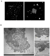
Detection of NiV N ordered structures with CLEM. NiV infected Vero cells were analyzed by fluorescence (A) and electron microscopy images (B,C) simultaneously. (A) NiV-infected cells, where NiV N is detected by spinning disc confocal microscopy. NiV ordered structures are indicated by white arrows. (B) The same syncytium as in image (A) is analyzed by electron microscopy. White arrows indicate NiV ordered structures. (C) The image on the right shows magnification of the NiV ordered clusters indicated with white arrows in images (A,B).
Figure 6
Figure 6
Solubility and three-dimensional analysis of NiV N, M, and P. On the left, solubility profiles of NiV N, NiV M, and NiV P are shown. The amino acid sequences of the NiV nucleoprotein N, the matrix protein M, and the phosphoprotein P were analyzed using the CamSol method in order to generate a solubility profile. Each amino acid in the protein sequence was assigned a score. Regions with scores higher than 1 denote highly soluble regions, while scores smaller than −1 indicate poor solubility. In the middle and on the right, three-dimensional (3D) representations of both: (i) the multimeric structure of NiV N, NiV M, and NiV P, with hydrophobic amino acids shown in red and non-hydrophobic amino-acids shown in blue, and (ii) the alignment of the NiV N, NiV M, and NiV P predicted monomeric structure (in magenta) and the monomeric template (in green) used in (i). The α-helices and β-sheets are indicated as ribbons and wide arrows, respectively.

Similar articles

Cited by

References

    1. Ang B.S.P., Lim T.C.C., Wang L. Nipah Virus Infection. J. Clin. Microbiol. 2018;56:e01875-17. doi: 10.1128/JCM.01875-17. - DOI - PMC - PubMed
    1. Hoenen T., Shabman R.S., Groseth A., Herwig A., Weber M., Schudt G., Dolnik O., Basler C.F., Becker S., Feldmann H. Inclusion bodies are a site of ebolavirus replication. J. Virol. 2012;86:11779–11788. doi: 10.1128/JVI.01525-12. - DOI - PMC - PubMed
    1. Dolnik O., Stevermann L., Kolesnikova L., Becker S. Marburg virus inclusions: A virus-induced microcompartment and interface to multivesicular bodies and the late endosomal compartment. Eur. J. Cell Biol. 2015;94:323–331. doi: 10.1016/j.ejcb.2015.05.006. - DOI - PubMed
    1. Heinrich B.S., Cureton D.K., Rahmeh A.A., Whelan S.P.J. Protein expression redirects vesicular stomatitis virus RNA synthesis to cytoplasmic inclusions. PLoS Pathog. 2010;6:e1000958. doi: 10.1371/journal.ppat.1000958. - DOI - PMC - PubMed
    1. Ringel M., Behner L., Heiner A., Sauerhering L., Maisner A. Replication of a Nipah Virus Encoding a Nuclear-Retained Matrix Protein. J. Infect. Dis. 2020;221((Suppl. 4)):S389–S394. doi: 10.1093/infdis/jiz440. - DOI - PubMed

Substances