Review
. 2022 Jul 19;14(7):1566.
doi: 10.3390/v14071566.

Hemagglutinin Subtype Specificity and Mechanisms of Highly Pathogenic Avian Influenza Virus Genesis

Affiliations
Review

Hemagglutinin Subtype Specificity and Mechanisms of Highly Pathogenic Avian Influenza Virus Genesis

Anja C M de Bruin et al. Viruses. .

Abstract

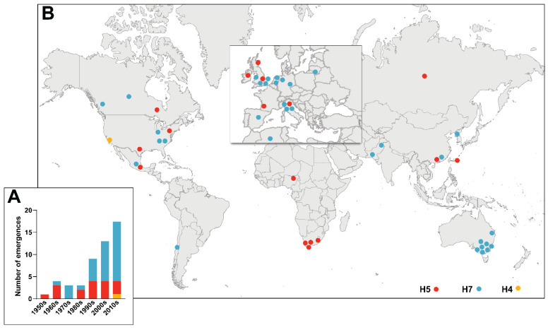
Highly Pathogenic Avian Influenza Viruses (HPAIVs) arise from low pathogenic precursors following spillover from wild waterfowl into poultry populations. The main virulence determinant of HPAIVs is the presence of a multi-basic cleavage site (MBCS) in the hemagglutinin (HA) glycoprotein. The MBCS allows for HA cleavage and, consequently, activation by ubiquitous proteases, which results in systemic dissemination in terrestrial poultry. Since 1959, 51 independent MBCS acquisition events have been documented, virtually all in HA from the H5 and H7 subtypes. In the present article, data from natural LPAIV to HPAIV conversions and experimental in vitro and in vivo studies were reviewed in order to compile recent advances in understanding HA cleavage efficiency, protease usage, and MBCS acquisition mechanisms. Finally, recent hypotheses that might explain the unique predisposition of the H5 and H7 HA sequences to obtain an MBCS in nature are discussed.

Keywords: Highly Pathogenic Avian Influenza Viruses; Low-Pathogenic Avian Influenza Viruses; RNA-dependent RNA polymerase; multibasic cleavage site; pathogen evolution; proteolytic cleavage; virulence.


Conflict of interest statement

The authors declare no conflict of interest.

Figures

Figure 1
Figure 1
Chronological and geographical depiction of the historical emergence of AIVs with MBCS 1959–2021. (A) Number of detections of novel AIVs with MBCS of the H5 (red), H7 (blue), and H4 (orange) HA subtypes per decade. (B) Geographical origin of MBCS-containing AIVs. Dots have been slightly displaced in areas with high emergence density in order to improve visibility.
Figure 2
Figure 2
Schematic representation and structure of the HA protein. (A) Schematic representation of HA following the cleavage of HA0 into the HA1 and HA2 subunits. The amino acids flanking the cleavage site (arrow head), fusion peptide, and the receptor binding and transmembrane domains are depicted. (B) Structure of H3 HA0 monomer (PDB entry 1HA0; non-cleaved R329Q mutant [108]), made in the PyMOL Molecular Graphics System version 2.5.2 Schrödinger LLC, showing the membrane-proximal cleavage loop and site (arrow head), and glycosylation site on position 22.
Figure 3
Figure 3
Alignment of cleavage site regions of all available newly emerged natural H5 and H7 MBCS-containing AIV sequences. Nucleotide and amino acid alignments of H5 MBCS-containing AIVs (A) and alignments of H7 MBCS-containing AIVs that did (B) or presumably did not (C) arise through NHR. The asterisks indicate strains that might have emerged through NHR based on codon usage and sequence alignment. The grey boxes delineate conserved amino acids from the LPAI consensus sequence with P1 to P4 indicated on top. Arginines and lysines are depicted in red and all other amino acids are depicted in blue. All sequences are available in fasta format as Supplementary Material.
Figure 4
Figure 4
Number of nucleotide substitutions necessary to acquire a tribasic cleavage site in HAs from LPAIVs. The number of single nucleotide substitutions required to obtain a tribasic cleavage site, defined as containing at least three basic amino acids in P1 to P4 and an arginine in P1, excluding histidine and pyrimidine-containing arginine codons based on codon usage in HPAIVs, in all reported LPAIV sequences from H1–H16 HA subtypes. The exact percent of sequences is indicated on top of each bar and a darker blue color indicates fewer substitutions required (Adapted from [183], reproduced with permission from Mathis Funk, Viruses; published by MDPI, 2022).
Figure 5
Figure 5
RNA secondary structures in the cleavage site region and their hypothesized influence on RdRp replication. (A) Examples of predicted cRNA structures encompassing the cleavage site region in H5 and H7 LPAIV and HPAIV. The predicted stem-loop (SL) structures are conserved in the H5 and H7 lineages. The nucleotides coding for (part of) the MBCS are inserted in the loop and depicted in red. The boundary between HA1 and HA2 is depicted with an arrowhead. Codons are distinguished from each other by dots (Adapted with permission from [219], available under the Creative Commons Attribution 4.0 International License). (B) Proposed model for increased stuttering and backtracking rates in the stem-loop region of HA due to RNA structure. The RdRp (PB1 in blue; PB2 in orange; PA in green) replicates the viral genome into product RNA (in red). Due to the close proximity of the template entry and exit channels in PB1, local structures based on complementary sequences in the template can form around the RdRp. The RdRp is trapped in the loop region, resulting in increased rates of stuttering and backtracking, leading to duplications in the RNA product.

References

    1. Stallknecht D.E., Shane S.M. Host range of avian influenza virus in free-living birds. Vet. Res. Commun. 1988;12:125–141. doi: 10.1007/BF00362792. - DOI - PubMed
    1. Olsen B., Munster V.J., Wallensten A., Waldenström J., Osterhaus A.D.M.E., Fouchier R.A.M. Global patterns of influenza A virus in wild birds. Science. 2006;312:384–388. doi: 10.1126/science.1122438. - DOI - PubMed
    1. Daoust P.Y., Kibenge F.S.B., Fouchier R.A.M., van de Bildt M.W.G., Van Riel D., Kuiken T. Replication of low pathogenic avian influenza virus in naturally infected mallard ducks (Anas platyrhynchos) causes no morphologic lesions. J. Wildl. Dis. 2011;47:401–409. doi: 10.7589/0090-3558-47.2.401. - DOI - PubMed
    1. Kuiken T. Is low pathogenic avian influenza virus virulent for wild waterbirds? Proc. R. Soc. B Biol. Sci. 2013;280:20130990. doi: 10.1098/rspb.2013.0990. - DOI - PMC - PubMed
    1. Webster R.G., Yakhno M., Hinshaw V.S., Bean W.J., Copal Murti K. Intestinal influenza: Replication and characterization of influenza viruses in ducks. Virology. 1978;84:268–278. doi: 10.1016/0042-6822(78)90247-7. - DOI - PMC - PubMed

Publication types

Substances